
INTERNATIONAL PROGRAMME ON CHEMICAL SAFETY
WORLD HEALTH ORGANIZATION
SUMMARY OF TOXICOLOGICAL DATA OF CERTAIN FOOD ADDITIVES
WHO FOOD ADDITIVES SERIES NO. 12
The data contained in this document were examined by the
Joint FAO/WHO Expert Committee on Food Additives*
Geneva, 18-27 April 1977
Food and Agriculture Organization of the United Nations
World Health Organization
* Twenty-first Report of the Joint FAO/WHO Expert Committee on Food
Additives, Geneva, 1977, WHO Technical Report Series No. 617
BROWN FK
EVALUATION FOR ACCEPTABLE DAILY INTAKE
BIOLOGICAL DATA
BIOCHEMICAL ASPECTS
After an intraperitoneal dose to a rat of 1.5 g/kg body weight
the extremities become orange in 60 minutes, and the animal sluggish.
The urine was normal. After 24 hours the animal was normal but urine
was deep orange-yellow (Goldblatt and Frodsham, 1952).
After an intraperitoneal dose to a rat of 1.5 g/kg bw the
extremeties become orange in 60 minutes, and the animal sluggish. The
urine was normal but urine was deep orange-yellow (Goldblatt and
Frodsham, 1952).
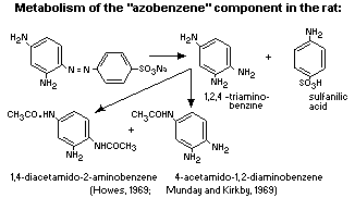
On incubation with the contents of rat ileum and caecum Brown FK
and its components undergo azo-reductive fission with formation of
sulfanilic acid, a phenazine-like material (P) and ill-defined
products that can be separated chrometographically. Brown FK, in
common with other brown azo coloutings, also undergoes azo-reductive
fission when incubated with rat-liver homogenate, but P has not been
detected among the products. Oral administration of Brown FK to rats,
pigs, rabbits and guinea-pigs results in the excretion of sulfanilic
acid in urine and faeces; P is detectable in trace/small amounts in
faeces, but is mainly present in caecal contents, predominantly during
the first six hours after dosing. A "blue material" is excreted in the
urine. On intraperitoneal administration to rats, Brown FK initially
gives rise to brown colouring in bile; later, sulfanilic acid and the
"blue material" appear in the urine. P is not found in faeces or in
caecal contents.
As formed in vitro from Brown FK, P is a complex mixture
comprising two main components P1 and P2. P1 has been identified as
1,4,7-tri-aminophenazine and P2 is probably a methylhomologue of P1
(Fore and Walker, 1967; Fore, Walker and Goldberg, 1967).
The metabolism of Brown FK would be extremely difficult to study
since the final metabolite mixture from the many components would be
exceedingly complex. Investigations have therefore been carried out on
the major components: 1,3-diamino-4-(p-sulfophenylazo)benzene
("azo-benzene" component) and 2,4-diamino-5-(p-sulfophenylazo)toluene
("azo-toluene" component). The metabolism of both components was
qualitatively similar, a proportion being excreted unchanged but the
bulk reductively cleaved to sulfanilic acid and the corresponding
amine (the latter being acetylated before excretion). Although the
metabolism of only these two components of Brown FK has been studied,
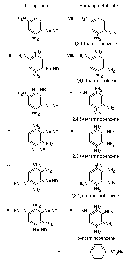 there is no reason to suppose that the metabolism of the other Brown
FK components should be fundamentally different. The primary metabolic
reaction in each case would be expected to be cleavage of the azo
linkages. A summary of the products of azo reduction of the six major
components of Brown FK is shown below (Howes, 1969; Munday and Kirkby,
1969).
there is no reason to suppose that the metabolism of the other Brown
FK components should be fundamentally different. The primary metabolic
reaction in each case would be expected to be cleavage of the azo
linkages. A summary of the products of azo reduction of the six major
components of Brown FK is shown below (Howes, 1969; Munday and Kirkby,
1969).
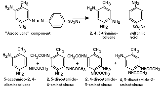
 Metabolism of the "azotoluene" component of Brown FK in the rat
Preliminary examnation of urine from rats fed the "azotoluene"
component of Brown FK showed the presence of sulfanilic acid and small
quantities of unchanged dye. Examination of an extract of this urine
revealed the presence of 5-acetamido-2,4-diaminoteluene (the major
metabolite), 2,5-diacetamido-4-aminotoluene, 2,4-diacetamido-5-amino-
toluene and 4,5-diacetamido-2-aminotoluene.
Unchanged dye was again identified in faecal extracts; no other
dye-derived compounds were detected.
The metabolism of the "azotoluene" component of Brown FK is
summarized below: (Munday 1969).
Metabolism of the "azotoluene" component of Brown FK in the rat
Preliminary examnation of urine from rats fed the "azotoluene"
component of Brown FK showed the presence of sulfanilic acid and small
quantities of unchanged dye. Examination of an extract of this urine
revealed the presence of 5-acetamido-2,4-diaminoteluene (the major
metabolite), 2,5-diacetamido-4-aminotoluene, 2,4-diacetamido-5-amino-
toluene and 4,5-diacetamido-2-aminotoluene.
Unchanged dye was again identified in faecal extracts; no other
dye-derived compounds were detected.
The metabolism of the "azotoluene" component of Brown FK is
summarized below: (Munday 1969).
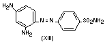
 An attempt was made to determine whether the "azobenzene"
component of Brown FK is reductively cleaved in humans, as in rats.
Reduction of the closely-related compound, protosil rubrum (XIII) has
been shown to take place in human subjects (Fuller, 1937).
An attempt was made to determine whether the "azobenzene"
component of Brown FK is reductively cleaved in humans, as in rats.
Reduction of the closely-related compound, protosil rubrum (XIII) has
been shown to take place in human subjects (Fuller, 1937).
 Administration of the "azobenzene" component of Brown FK to human
subjects led to no detectable unchanged dye in the urine, and no
appreciable urinary sulfanilic acid. It can be inferred from these
results that the "azobenzene" component is not absorbed from the
intestine as such, but they give no information on the possible
reduction of this compound in vivo, since it was shown that orally
administered sulfanilic acid was not absorbed in man. Sulfanilic acid,
if formed from the dye, would therefore be excreted in the faeces; the
experimental confirmation of this would be technically very difficult
and these studies were not pursued further (Jenkins and Favell, 1971).
1,2,4-triaminobenzene and 2,4,5-triaminotoluene have been shown
to uncouple oxidative phosphorylation in vitro, interfering with ATP
production in the muscle cell and to ionic imbalance with cell death
(Munday, 1971).
Acute toxicity
Animal Route LD50 mg/kg body weight Reference
Mouse oral >2 000 (with salt) Grasso et al., 1968
oral 1 100-2 250 (with salt) Edwards and Wilson, 1966
oral 960-1 140 (no salt) Edwards and Wilson, 1966
i.p. 1 500-2 000 (with salt) Grasso et al., 1968
i.p. 960-1 720 (with salt) Edwards and Wilson, 1966
i.p. 840- 880 (no salt) Edwards and Wilson, 1966
Rat oral > 8 000 (with salt) Grasso et al., 1968
oral 900-1 910 (with salt) Edwards and Wilson, 1966
oral 780- 970 (no salt) Edwards and Wilson, 1966
i.p. 750-1 150 (with salt) Grasso et al., 1968
i.p. 1 100-2 250 (with salt) Edwards and Wilson, 1966
i.p. 960-1 150 (no salt) Edwards and Wilson, 1966
Guinea-pig oral 3 000 (with salt) Edwards and Wilson, 1966
oral 2 610 (no salt) Edwards and Wilson, 1966
i.p. 900 (with salt) Edwards and wilson, 1966
i.p. 780 (no salt) Edwards and Wilson, 1966
Rabbit oral 450- 680 (with salt) Edwards and Wilson, 1966
oral 390- 590 (no salt) Edwards and Wilson, 1966
Chicken oral > 10, 000 (with salt) Edwards and Wilson, 1966
oral >8 700 (no salt) Edwards and Wilson, 1966
For all species, animals dying did so from within a few minutes
to 96 hours. Many animals, after either oral or intraperitoneal
treatment, showed lack of coordination, hypersensitivity and
hyperactivity; convulsions usually preceded death (Edwards and Wilson,
1966).
Meningeal congestion or haemorrhage was seen at post-mortem
examination in rats and mice which died following both oral and
intraperitoneal treatment with 3.4 g/kg of Brown FK as a 10% solution.
This was the highest dose administered and the condition may have been
present to a lesser degree in animals treated with lower levels of
Brown FK but postmortem identification of the lesion was made
difficult by tissue coloration. The meningeal congestion/haemorrhage
was probably caused by the sodium chloride in the dye solution since
we have observed this lesion after administration of hypertonic
solutions of sodium chloride to rats. In this instance, the lowest
dose levels at which the meningeal lesion has been observed were
6.0 g/kg of sodium chloride orally as a 10% solution and 4.0 g/kg
intraperitoneally as a 5% solution (Edwards and Wilson, 1966).
Following oral intubation, external tissue coloration was
apparent after some hours in rats and guinea-pigs. No coloration of
the tissues was seen in rabbits and chickens. After intraperitoneal
injection, external tissue coloration was apparent and intense after a
few minutes in the rats and guinea-pigs.
Colour was seen in the faeces of rats, mice, rabbits and guinea-
pigs up to 24 hours a£ter oral treatment; it was also excreted in the
urine of rats, mice, guinea-pigs and rabbits within 15 minutes of
either oral or intraperitoneal treatment (Edwards and Wilson, 1966).
Hearts from some rats and mice surviving for 21 days after
treatmant were examined histologically. A degenerative lesion was
found in 15% of rats given orally 1-2.5 g/kg body weight but not with
3.37 g/kg. The same lesions were found in mice in 50% given orally
0.9 g/kg but not if given 0.6, 1.35 and 2.03 g/kg. If given
intraperitoneal 25-60% of mice showed lesions at 0.75 and 1.03 g/kg
body weight (Edwards and Wilson, 1966).
Amines derived from Brown FK and from its two myotoxie
components, 2,4-diamino-5-(p-sulfophenylazo)toluene and 1,3-diamino-4-
(p-sulfo-phenylazo)benzene were injected intravenously into rats in
single doses of 3.13-25 mg/kg. The mixture of amines from Brown FK was
also injected into mice in the same range of doses. Cardiac and
muscular lesions were produced by the amines in both species. These
amines are biological degradation products in the intestine. The
finding that orally administered Brown FK is myotoxic in rats but not
in mice is probably due to differences in the intestinal flora in the
two species (Walker, Grasso and Gaunt, 1970).
Investigation of the pigment deposited
After feeding Brown FK to rats and mice pigment is found in
heart, skeletal muscle, tongue, diaphragm, thyroids, brain, liver,
kidneys, spleen, lungs, pancreas, bladder, testes, ovary, uterus,
skin, stomach, duodenum, ileum, brown fat and bone marrow. In
addition, a pigment has been detected in the plasma of rats.
Staining tests commonly used to identify lipofuscin were negative
with the exception of the test for metachromasia with toluidine blue.
The tests applied were as follows:
Usual response of Response of Brown FK
known lipofuscin induced pigment
Test for iron negative negative
Sudan fat stains positive negative
Reduction of
ferric salts positive negative
Reduction of
ammoniacal silver
salts positive negative
Basophilic properties positive negative
Periodic acid -
schiff reaction positive negative
Acid fastness acid fast negative
Toluidine blue
at pH 3 stains greenish
metachromatically
green
Two further histochemical tests clearly differentiate between
lipo-fuscin and the Brown FK - induced pigment:
- Potassium permanganate/oxalic acid bleached lipofuscin, but not
the brown FK - induced pigment.
- Sodium dithionite bleached both lipofusein and the Brown FK
induced pigment. However, after rinsing and allowing to stand in air,
the Brown FK - induced pigment reappeared; lipofusein was permanently
bleached.
The Brown FK - induced pigment does not fluoresce in ultra-violet
light. All the samples of lipofuscin which we have examined were
fluorescent. Pigment has been found in the thyroid, brown fat and bone
marrow; these tissues have not been recorded as being sites for
lipofuscin deposition. Furthermore, a coloured substance has been
demonstrated in the plasma, never lipofuscin.
The speed at which the Brown FK - induced pigment is deposited is
uncharacteristic of lipofuscin information. In acute studies, pigment
has been seen in the intestinal wall and villi within 24 hours of
feeding the dye, and in the kidney after five days. Pigment masses
produced in macrophages either in vivo after the intraperitoneal
injection of Brown FK into mice, or in vitro, when Brown FK was
incorporated in the macro-phage culture medium, appeared identical.
Tests for lipofuscin proved negative; the pigment in the macrophages
closely resembled that seen in macrophages in stained sections of
tissues taken from rats and mice fed Brown FK.
Electron microscope studies have identified differences in
morphology between lipofuscin and the Brown FK - induced pigment. In
aged rats and mice fed Brown FK, conjugate forms were observed in
which induced pigment and control lysosomal material appeared in the
same membrane limited body (Hope, 1971).
It is likely that this compound oxidises within the cell to
1,4,7-triaminophenazine
Administration of the "azobenzene" component of Brown FK to human
subjects led to no detectable unchanged dye in the urine, and no
appreciable urinary sulfanilic acid. It can be inferred from these
results that the "azobenzene" component is not absorbed from the
intestine as such, but they give no information on the possible
reduction of this compound in vivo, since it was shown that orally
administered sulfanilic acid was not absorbed in man. Sulfanilic acid,
if formed from the dye, would therefore be excreted in the faeces; the
experimental confirmation of this would be technically very difficult
and these studies were not pursued further (Jenkins and Favell, 1971).
1,2,4-triaminobenzene and 2,4,5-triaminotoluene have been shown
to uncouple oxidative phosphorylation in vitro, interfering with ATP
production in the muscle cell and to ionic imbalance with cell death
(Munday, 1971).
Acute toxicity
Animal Route LD50 mg/kg body weight Reference
Mouse oral >2 000 (with salt) Grasso et al., 1968
oral 1 100-2 250 (with salt) Edwards and Wilson, 1966
oral 960-1 140 (no salt) Edwards and Wilson, 1966
i.p. 1 500-2 000 (with salt) Grasso et al., 1968
i.p. 960-1 720 (with salt) Edwards and Wilson, 1966
i.p. 840- 880 (no salt) Edwards and Wilson, 1966
Rat oral > 8 000 (with salt) Grasso et al., 1968
oral 900-1 910 (with salt) Edwards and Wilson, 1966
oral 780- 970 (no salt) Edwards and Wilson, 1966
i.p. 750-1 150 (with salt) Grasso et al., 1968
i.p. 1 100-2 250 (with salt) Edwards and Wilson, 1966
i.p. 960-1 150 (no salt) Edwards and Wilson, 1966
Guinea-pig oral 3 000 (with salt) Edwards and Wilson, 1966
oral 2 610 (no salt) Edwards and Wilson, 1966
i.p. 900 (with salt) Edwards and wilson, 1966
i.p. 780 (no salt) Edwards and Wilson, 1966
Rabbit oral 450- 680 (with salt) Edwards and Wilson, 1966
oral 390- 590 (no salt) Edwards and Wilson, 1966
Chicken oral > 10, 000 (with salt) Edwards and Wilson, 1966
oral >8 700 (no salt) Edwards and Wilson, 1966
For all species, animals dying did so from within a few minutes
to 96 hours. Many animals, after either oral or intraperitoneal
treatment, showed lack of coordination, hypersensitivity and
hyperactivity; convulsions usually preceded death (Edwards and Wilson,
1966).
Meningeal congestion or haemorrhage was seen at post-mortem
examination in rats and mice which died following both oral and
intraperitoneal treatment with 3.4 g/kg of Brown FK as a 10% solution.
This was the highest dose administered and the condition may have been
present to a lesser degree in animals treated with lower levels of
Brown FK but postmortem identification of the lesion was made
difficult by tissue coloration. The meningeal congestion/haemorrhage
was probably caused by the sodium chloride in the dye solution since
we have observed this lesion after administration of hypertonic
solutions of sodium chloride to rats. In this instance, the lowest
dose levels at which the meningeal lesion has been observed were
6.0 g/kg of sodium chloride orally as a 10% solution and 4.0 g/kg
intraperitoneally as a 5% solution (Edwards and Wilson, 1966).
Following oral intubation, external tissue coloration was
apparent after some hours in rats and guinea-pigs. No coloration of
the tissues was seen in rabbits and chickens. After intraperitoneal
injection, external tissue coloration was apparent and intense after a
few minutes in the rats and guinea-pigs.
Colour was seen in the faeces of rats, mice, rabbits and guinea-
pigs up to 24 hours a£ter oral treatment; it was also excreted in the
urine of rats, mice, guinea-pigs and rabbits within 15 minutes of
either oral or intraperitoneal treatment (Edwards and Wilson, 1966).
Hearts from some rats and mice surviving for 21 days after
treatmant were examined histologically. A degenerative lesion was
found in 15% of rats given orally 1-2.5 g/kg body weight but not with
3.37 g/kg. The same lesions were found in mice in 50% given orally
0.9 g/kg but not if given 0.6, 1.35 and 2.03 g/kg. If given
intraperitoneal 25-60% of mice showed lesions at 0.75 and 1.03 g/kg
body weight (Edwards and Wilson, 1966).
Amines derived from Brown FK and from its two myotoxie
components, 2,4-diamino-5-(p-sulfophenylazo)toluene and 1,3-diamino-4-
(p-sulfo-phenylazo)benzene were injected intravenously into rats in
single doses of 3.13-25 mg/kg. The mixture of amines from Brown FK was
also injected into mice in the same range of doses. Cardiac and
muscular lesions were produced by the amines in both species. These
amines are biological degradation products in the intestine. The
finding that orally administered Brown FK is myotoxic in rats but not
in mice is probably due to differences in the intestinal flora in the
two species (Walker, Grasso and Gaunt, 1970).
Investigation of the pigment deposited
After feeding Brown FK to rats and mice pigment is found in
heart, skeletal muscle, tongue, diaphragm, thyroids, brain, liver,
kidneys, spleen, lungs, pancreas, bladder, testes, ovary, uterus,
skin, stomach, duodenum, ileum, brown fat and bone marrow. In
addition, a pigment has been detected in the plasma of rats.
Staining tests commonly used to identify lipofuscin were negative
with the exception of the test for metachromasia with toluidine blue.
The tests applied were as follows:
Usual response of Response of Brown FK
known lipofuscin induced pigment
Test for iron negative negative
Sudan fat stains positive negative
Reduction of
ferric salts positive negative
Reduction of
ammoniacal silver
salts positive negative
Basophilic properties positive negative
Periodic acid -
schiff reaction positive negative
Acid fastness acid fast negative
Toluidine blue
at pH 3 stains greenish
metachromatically
green
Two further histochemical tests clearly differentiate between
lipo-fuscin and the Brown FK - induced pigment:
- Potassium permanganate/oxalic acid bleached lipofuscin, but not
the brown FK - induced pigment.
- Sodium dithionite bleached both lipofusein and the Brown FK
induced pigment. However, after rinsing and allowing to stand in air,
the Brown FK - induced pigment reappeared; lipofusein was permanently
bleached.
The Brown FK - induced pigment does not fluoresce in ultra-violet
light. All the samples of lipofuscin which we have examined were
fluorescent. Pigment has been found in the thyroid, brown fat and bone
marrow; these tissues have not been recorded as being sites for
lipofuscin deposition. Furthermore, a coloured substance has been
demonstrated in the plasma, never lipofuscin.
The speed at which the Brown FK - induced pigment is deposited is
uncharacteristic of lipofuscin information. In acute studies, pigment
has been seen in the intestinal wall and villi within 24 hours of
feeding the dye, and in the kidney after five days. Pigment masses
produced in macrophages either in vivo after the intraperitoneal
injection of Brown FK into mice, or in vitro, when Brown FK was
incorporated in the macro-phage culture medium, appeared identical.
Tests for lipofuscin proved negative; the pigment in the macrophages
closely resembled that seen in macrophages in stained sections of
tissues taken from rats and mice fed Brown FK.
Electron microscope studies have identified differences in
morphology between lipofuscin and the Brown FK - induced pigment. In
aged rats and mice fed Brown FK, conjugate forms were observed in
which induced pigment and control lysosomal material appeared in the
same membrane limited body (Hope, 1971).
It is likely that this compound oxidises within the cell to
1,4,7-triaminophenazine
 This would also explain the behaviour of the pigment with
sodium dithio-nite. The latter reduces the phenazine ring to
5,10-dihydroderivative which is probably colourless. After exposure to
air reoxidation would occur. Thus the pigment may not represent
evidence of sub-lethal cell damage, but is more an insoluble oxidation
product of a dye metabolite.
1,2,4-triaminobenzene was very rapidly oxidized to
1,4,7-triamino-phenazine by a mitochondrial suspension; no
phenazine derivatives were detected with triaminotoluene under
the same circumstances (Kirby, 1968).
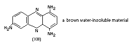
1,4,7-triaminophenazine is a brown, water-insoluble material,
which is very readily formed from 1,2,4-triaminobenzene (Muller,
1889).
Short-term studies
Mouse
Groups of 10 male and 10 female mice received the colour (both
fresh and stored) at the level of 1 g/kg daily for three weeks. A
significant reduction in weight gain was noted in the mice receiving
the stored solution but not in those receiving fresh solution. One
male and one female receiving the fresh solution showed cardiac
lesions (BIBRA, 1964).
Daily oral or intraperitoneal doses up to 2 g/kg or 1 g/kg
respectively for 43 days to groups of 10 or 12 mice were well
tolerated (Grasso et al., 1968).
Groups of 10 male and 10 female mice (Colworth C57 B1 strain,
initially six weeks old) were fed for 90 days on a synthetic diet
containing 0, 0.05, 0.075, 0.10, 0.25, 0.50, 0.75, 1.0 and 2.0% of
Brown FK (equivalent to 0, 0.025, 0.0375, 0.05, 0.125, 0.25, 0.375,
0.50 and 1% Brown FK coloured components) composition 51% colour,
47% salt. A further group of 20 mice were fed synthetic diet
containing added 1.0% sodium chloride as a control for the additional
dietary salt derived from the Brown FK. At the 0.125% dietary colour
level pigment deposition occurred in tissues. At 0.25% and above there
was splenic enlargement, at 0.5% liver and heart were also enlarged
and at 1% there was reduced growth, poor food utilization, liver,
spleen, heart and testicular enlargement and histological evidence of
degenerative heart lesions. Thyroids, muscle, intestine and squamous
part of stomach were pigmented (Ashmole et al., 1958).
Rat
Groups of animals received the colour at the level of 0.5 g
Brown FK per kg body weight for three weeks, orally or
intraperitoneally. When dosed orally, 20 rats were treated and six
animals died between five and 11 doses. Post-mortem examination of
rats dying during the test or killed at the end revealed general
tissue staining in five rats. Of 18 hearts examined histologically,
eight showed degenerative lesions and a brown pigment was observed in
small amounts in nine hearts after three weeks.
In the multiple dose intraperitoneal test, eight rats were
treated and none died during the treatment period. General organ
staining was observed in all animals at post-mortem examination.
Hearts from seven rats were examined microscopically and degenerative
lesions were found in one heart and small amounts of pigment in three
hearts after three weeks (Kirkby, 1968).
No ill-effects were seen in three weanling rats given a 0.1%
solution for 28 days, the intake being 15 mg/day (Goldblatt and
Frodsham, 1952).
Administration of two or three oral doses of 1 g/Brown FK/kg bw
to rats induced a myopathy in cardiac and skeletal muscles
characterized by multiple vacuoles about 1-2 µ in diameters.
Ultrastructurally, these were shown to consist of areas of
fibrillolysis. Histochemically, the myopathy was accompanied by a
moderate increase in acid-phosphatase activity and by a loss of
phosphorylase activity. Subsequently complete lysis of the affected
fibres ensued. In the heart, lysis was followed by macrophage invasion
and fibroblastic proliferation, and in skeletal muscle by
regeneration. The occurrence of lipofuscin in muscle fibres and in
macrophages was scanty and erratic. When Brown FK was given in the
diet at a level of 2%, fibrillolysis and an increase in the number and
electron-density of lysosomes was observed ultrastructurally during
week two to three of the test. These changes were accompanied by a
marked elevation of histochemically demonstrable acid phosphatase.
Progressive deposition of lipofuscin was the principal pathological
feature during week three to 12 (Grasso et al, 1968).
Daily oral doses up to 2 g/kg for 43 days to groups of 10 rats
induced rapid loss of weight and death, with severe damage to cardiac
and skeletal muscle, characterized by vacuolar myopathy and lipofuscin
deposition. Of three pure components of Brown FK studied, one
(2,4-diamino-5-(p-sulfophenylazo)toluene) and to a lesser extent
another (1,3-diamino-4-(p-sulfophenylazo)benzene) produced similar,
but not identical lesions to those induced by the parent colouring
after repeated oral doses of 0.5 g/kg. Ultrastructural studies
confirmed the extensive loss of myofibrillar elements and
histochemical studies revealed a loss in the activity of mitochondrial
enzymes. Similar intraperitoneal injections in doses up to 1.0 g/kg
for 43 days to groups of 10 or 12 rats did not have any effect on the
heart or skeletal muscle (Grasso et al., 1968).
In a series of experiments using groups of 10-12 rats receiving
the colour at levels of 1 and 0.1 g/kg orally and 1, 0.25 and 0.1 g/kg
intraperitoneally daily up to a maximum of 43 doses the following
observations were made. A specific cardiac lesion was identified at
the oral dose of 1 g/kg. There were large areas of myocardial necrosis
and replacement by large mononuclears, with involvement of the
sub-pericardial region and endocardium. Some myocardial cells had lost
their stainable cytoplasm and appeared only as empty sheaths.
Intraperitoneally, the colour produced little or no cardiac damage at
any dose tested. At these high doses of 1 g/kg, most animals showed
congestion, fatty change or necrosis of the liver with hydropic
degeneration of the kidney. There was no obvious splenomegaly. Daily
doses of 100 mg/kg by stomach tube produced two pericardial and one
sub-pericardial lesions. In addition, early hydropic degeneration of
the kidney was seen in two rats with one of those animals also showing
fatty change in the liver (BIBRA, 1964).
Administration of Brown FK (purity 80.0%) at dietary levels of
0, 0.001, 0.01, 0.1 and 1.0% for 150 days showed no adverse effects on
growth, food consumption, haematological indices, liver and kidney
function and organ weights. One male rat at the 1.0% level showed the
typical myocardial changes, other rats showed deposits of lipofuscin
especially in females. The no-effect level was 0.1% (Gaunt et al.,
1968).
Groups of 12 male and 12 female rats (Colworth Wistar strain,
initially three to four weeks old) were fed for 112 days on a
commercial stock diet, containing 0, 0.05, 0.1, 0.5, 1.0 and 2.0% of
Brown FK (0, 0.025, 0.05, 0.25, 0.5 and 1% Brown FK coloured
components) 51% dye component, 47% salt. A further group of 24 rats
were fed the commercial stock containing an added 1% sodium chloride
as a control for the additional dietary salt derived from the
Brown FK.
In addition, to eliminate damage to the heart from cardiac
puncture in any rat kept to 16 weeks, groups of six male and six
female rats were fed for six weeks on powdered stock diet containing
0, 0.05, 0.5 and 2.0% of Brown FK (0, 0.025, 0.25 and 1.0% Brown FK
coloured components). A group of 12 rats also received 1.0% sodium
chloride added to the basic diet. All these rats were used for
biochemical tests during weeks 0-6 after which they were killed. At
the 0.25% level tissue pigmentation appeared, at 0.5% liver
enlargement occurred and at the 1% level there was reduced growth,
poor food utilization, enlargement of liver, testes and thyroid,
histological evidence of degenerative heart lesions, increased urinary
indican excretion and elevation of SGOT. The intestine and squamous
portion of stomach as well as thyroid were stained. The no effect
level was 0.05% (Ashmole et al., 1966).
Pig
Groups of female and male pigs were given doses of 0, 100, 250
and 500 mg/kg/day for 24 weeks without adverse effects on growth, food
consumption, haematological indices, liver and kidney function and
organ weights. Lipofuscin was widely distributed in both sexes at all
dose levels in one or more organs. It particularly affected the liver,
where it was accompanied by increased lysosomal enzyme activity, more
marked at the higher dose levels. It was also seen in the heart in
males and here it was associated with an increased acid-phosphatase
activity, and in the kidneys, at the highest dose level in females and
at all levels in males. A no-effect level was not seen (Gaunt et al.,
1968).
Special studies on "azobenzene" and "azotoluene" components
Mouse
Groups of three male and three female mice (C57 B1) were fed 0,
0.5 and 1.0% of components in a synthetic type diet for six weeks.
With both compounds the thyroids were dark and intestine and squamous
portion of stomach were stained salmon pink. Heart lesions were seen
in all mice fed 1% azotoluene and not in those given azobenzene. More
pigment was seen in mice fed azobenzene component, little in those fed
azo-toluene component (Kirkby, 1968).
Rat
Groups of three male and three female Colworth Wistar rats were
fed azobenzene and azotoluene component at dietary levels of 0, 0.5
and 1% in commercial stock diet for six weeks. The thyroids of rats on
"azo-benzene" were dark, heart, muscle and brain were stained but the
intestine was stained only slightly. Pale hearts and meningeal
haemorrhage were seen with "azotoluene", otherwise pigmentation was as
with "azobenzene". One-sixth "azobenzene" and 4/5 "azotoluene" rats
had heart lesion. Most pigment was seen histologically in "azobenzene"
rats, least in "azo-toluene" rats (Kirkby, 1968).
In another study 1,2,4-triaminobenzene was given to groups of six
to seven rats orally five days per week for two weeks at 50, 60, 75
and 100 mg/kg body weight/day. Six-sevenths receiving 100 mg/kg/day
died after three doses with severe heart lesions, 7/11 on 75 mg/kg/day
also died after three doses. Heart pigmentation occurred after five
days treatment or longer. Animals on lower doses showed both extensive
heart pigmentation and cardiac necrosis. 1,2,4,5-tetraaminobenzene was
given to groups of six rats orally five days per week for two weeks at
150 and 200 mg/kg body weight/day. 1,2,3,4-tetraaminobenzene was given
to groups of six rats orally five days per week for two weeks at 125
and 166 mg/kg body weight/day. No frank heart lesions and only
instances of diffuse increase in interstitial cells in the heart were
observed. No heart or thyroid pigment deposition was seen (Mulky et
al., 1969).
Long-term studies
Mouse
Groups of 40 male and 40 female Colworth C57 B1 mice were fed for
80 weeks on a synthetic diet containing 0, 0.0125%, 0.0375%, 0.075%,
0.125% and 0.625% Brown FK coloured components (Brown FK purchased
contained 62.5% coloured components). Only at the 0.625% was there
reduced growth and food utilization and higher mortality among
females. There was increased liver, kidney, spleen, brain and testes
weight, evidence of splenic haemopoiesis, increased myocardial
fibrosis. Heart weight was increased at the 0.125% level. Increased
hepatic nodules were seen as from 0.075% and pigment deposition as
from 0.0375% level. At termination, after 80 weeks, the number of
animals with nodules for the different dose levels was 26, 23, 27, 56,
42 and 64 respectively. Increased hepatic nodules were observed at
dose level 0.075% and higher. The number of mice with hepatocellular
carcinoma were 3, 2, 0, 5, 6 and 2 respectively. Pigment deposition
was observed at dose level 0.0375% and higher (Wilson et al., 1970).
Rat
Groups of 32 male and 36 female Colworth Wistar rats were fed for
two years on a synthetic diet containing 0, 0.01%, 0.03%, 0.06%, 0.1%
and 0.5% of Brown FK coloured components (Brown FK purchased contained
54.2% coloured components). Only at the 0.5% level was there increased
splenic weight and hepatic granulomata. Pigment deposition was seen as
from 0.06%. The no-effect level for pigment deposition was 0.03% and
0.06% when based on toxicity evidence (Wilson et al., 1971).
Special studies on mutagenicity
Brown FK and its constituents were assayed for mutagenicity in
Salmonella typhimurium TA 1535, TA 1537 and TA 1538 when activated
by a rat liver supernatant fraction. Mutagenicity was linearly dose-
dependent in the range 0-3 mg/plate with activities ranging from 22 to
50 times the spontaneous mutation frequency. One sample of Brown FK
was mutagenic in the absence of metabolic activation producing a
16-fold increase in mutation at 4 mg/plate. Two major constituents of
Brown FK, 2,4-diamino-5-(p-sulfophenylazo)toluene (I) and 1,3-diamino-
4-(p-sulfo-phenylazo)benzene (II) each present at about 18% in the
complete colour, were mutagenic in TA 1538. Mutagenicity was
linearly dose-related in the range 0-1 µmol/plate, with slopes of
0.35 mutants/nmol for compound I and 1.5 mutants/nmol for compound II.
This activity was dependent on metabolic activation. Four other major
constituents were inactive, as was sulfanilic acid, the major
excretion product. The mutagenicity of Brown FK could be largely
accounted for by the combined effects of compound I and II (Venitt and
Bushell, 1976).
REFERENCES
Ashmole, R. T., Campbell, P., Kirkby, W. W. and Wilson, R. (1966)
Effects of feeding dietary Brown FK to rats for six and 16 weeks.
Unpublished report from Unilever Research Laboratories, submitted to
the World Health Organization by Unilever Ltd.
Ashmole, R. T., Kirkby, W. W. and Wilson, R. (1958) Thirteen week
mouse feeding trial. Unpublished report from Unilever Research
Laboratories, submitted to the World Health Organization by Unilever
Ltd.
Edwards, K, B. and Wilson, R. (1966) Acute toxicity of Brown FK in
rats, mice, guinea-pigs, rabbits and chickens. Unpublished report from
Unilever Research Laboratories
Fore, H. and Walker, R, (1967) Studies on Brown FK. I. Composition and
synthesis of components, Fd. and Cosmet. Toxicol., 5, 1-9
Fore, H., Walker, R. and Golberg (1967) Studies on Brown FK. II.
Degradative changes undergone in vitro and in vivo, Fd. and
Cosmet. Toxicol., 5, 459-473
Fuller, A. T. (1937) Lancet, 194
Gaunt, I. F., Hall, D. E., Grasso, P. and Golberg, L. (1968) Studies
on Brown FK. V. Shortterm feeding studies in the rat and pig,
Fd. and Cosmet. Toxicol., 6, 301-312
Goldblatt and Frodsham (1952) Private communication from ICI
(unpublished report)
Grasso, P., Muir, A., Golberg, L. and Batstone, E. (1968) Cytopathic
effects of Brown FK on cardiac and skeletal muscle in the rat,
Fd. and Cosmet. Toxicol., 6, 13-24
Grasso, P., Gaunt, I. F., Hall, D. E., Golherg, L. and Batstone, E.
(1968) Studies on Brown FK. III. Administration of high doses to rats
and mice, Fd. and Cosmet. Toxicol., 6, 1-11
Hope, J. (1971) Ultrastructure of the pigment induced in various
tissues of the rat by long-term feeding of the dye Brown FK.
Unpublished report from Unilever Research Laboratories, submitted to
the World Health Organization by Unilever Ltd.
Howes, D. (1969) Metabolism of 14C labelled 1,3-diamino-4-
(p-sulpho-phenylazo) benzene, a component of the dye Brown FK, in the
rat, Unpublished report from Unilever Research Laboratories, submitted
to the World Health Organization by Unilever Ltd.
Jenkins, F. P. and Favell, D. J. (1971) Metabolism of the
"monoazobenzene" component of Brown FK in human subjects. Unpublished
report from Unilever Research Laboratories, submitted to the World
Health Organization by Unilever Ltd.
Kirkby, W. W. (1968) Effects of Brown FK and two of its constituents
on pigment deposition and lesions in rats and mice. Unpublished report
from Unilever Research Laboratories, submitted to the World Health
Organization by Unilever Ltd.
Kirkby, W. W. (1968) Nature of the pigment induced in tissues of rats
and mice fed Brown FK. Unpublished report from Unilever Research
Laboratories, submitted to the World Health Organization by Unilever
Ltd.
Mulky, M. J., Munday, R., Ashmole, R. T. and Kirkby, W. W. (1969)
Evaluation of the terminal causative agent in Brown FK induced
myopathy and pigment deposition. Unpublished report from Unilever
Research Laboratories, submitted to the World Health Organization by
Unilever Ltd.
Muller, E. (1889) Chem. Ber., 22, 856
Munday, R. (1969) Metabolism of 2,4-diamino-5-(p-sulphophenylazo)
toluene. Unpublished report from Unilever Research Laboratories,
submitted to the World Health Organization by Unilever Ltd.
Munday, R. (1971) Uncoupling of oxidative phosphorylation by Brown FK
metabolites. Unpublished report from Unilever Research Laboratories,
submitted to the World Health Organization by Unilever Ltd.
Munday, R. and Kirkby, W. W. (1969) Metabolism of 1,3-diamino-4-
(p-sulpho-phenylazo) benzene. Unpublished report from Unilever
Research Laboratories, submitted to the World Health Organization by
Unilever Ltd.
Venitt, S. and Bushell, 0. T. (1976) Mutagenicity of the food colour
Brown FK and constituents in Salmonella typhimurium, Mutation
Research, 40, 309-316
Walker, R., Grasso, P. and Gaunt, I. F. (1970) Myotoxtcity of amine
metabolites from Brown FK, Fd. and Cosmet Toxicol., 8, 539-542
Wilson, R., Gellatly, J. B. M., Kirkby, W. W. and Ashmole, R. T.
(1970) Biological evaluation of Brown FK: 80-week mouse feeding trial.
Unpublished report from Unilever Research Laboratories, submitted to
the World Health Organization by Unilever Ltd.
Wilson, R., Gellatly, J. B. M., Kirkby, W. W. and Ashmole, R. T.
(1971) Biological evaluation of Brown FK: 2-year rat feeding trial.
Unpublished report from Unilever Research Laboratories, submitted to
the World Health Organization by Unilever Ltd.
This would also explain the behaviour of the pigment with
sodium dithio-nite. The latter reduces the phenazine ring to
5,10-dihydroderivative which is probably colourless. After exposure to
air reoxidation would occur. Thus the pigment may not represent
evidence of sub-lethal cell damage, but is more an insoluble oxidation
product of a dye metabolite.
1,2,4-triaminobenzene was very rapidly oxidized to
1,4,7-triamino-phenazine by a mitochondrial suspension; no
phenazine derivatives were detected with triaminotoluene under
the same circumstances (Kirby, 1968).
1,4,7-triaminophenazine is a brown, water-insoluble material,
which is very readily formed from 1,2,4-triaminobenzene (Muller,
1889).
Short-term studies
Mouse
Groups of 10 male and 10 female mice received the colour (both
fresh and stored) at the level of 1 g/kg daily for three weeks. A
significant reduction in weight gain was noted in the mice receiving
the stored solution but not in those receiving fresh solution. One
male and one female receiving the fresh solution showed cardiac
lesions (BIBRA, 1964).
Daily oral or intraperitoneal doses up to 2 g/kg or 1 g/kg
respectively for 43 days to groups of 10 or 12 mice were well
tolerated (Grasso et al., 1968).
Groups of 10 male and 10 female mice (Colworth C57 B1 strain,
initially six weeks old) were fed for 90 days on a synthetic diet
containing 0, 0.05, 0.075, 0.10, 0.25, 0.50, 0.75, 1.0 and 2.0% of
Brown FK (equivalent to 0, 0.025, 0.0375, 0.05, 0.125, 0.25, 0.375,
0.50 and 1% Brown FK coloured components) composition 51% colour,
47% salt. A further group of 20 mice were fed synthetic diet
containing added 1.0% sodium chloride as a control for the additional
dietary salt derived from the Brown FK. At the 0.125% dietary colour
level pigment deposition occurred in tissues. At 0.25% and above there
was splenic enlargement, at 0.5% liver and heart were also enlarged
and at 1% there was reduced growth, poor food utilization, liver,
spleen, heart and testicular enlargement and histological evidence of
degenerative heart lesions. Thyroids, muscle, intestine and squamous
part of stomach were pigmented (Ashmole et al., 1958).
Rat
Groups of animals received the colour at the level of 0.5 g
Brown FK per kg body weight for three weeks, orally or
intraperitoneally. When dosed orally, 20 rats were treated and six
animals died between five and 11 doses. Post-mortem examination of
rats dying during the test or killed at the end revealed general
tissue staining in five rats. Of 18 hearts examined histologically,
eight showed degenerative lesions and a brown pigment was observed in
small amounts in nine hearts after three weeks.
In the multiple dose intraperitoneal test, eight rats were
treated and none died during the treatment period. General organ
staining was observed in all animals at post-mortem examination.
Hearts from seven rats were examined microscopically and degenerative
lesions were found in one heart and small amounts of pigment in three
hearts after three weeks (Kirkby, 1968).
No ill-effects were seen in three weanling rats given a 0.1%
solution for 28 days, the intake being 15 mg/day (Goldblatt and
Frodsham, 1952).
Administration of two or three oral doses of 1 g/Brown FK/kg bw
to rats induced a myopathy in cardiac and skeletal muscles
characterized by multiple vacuoles about 1-2 µ in diameters.
Ultrastructurally, these were shown to consist of areas of
fibrillolysis. Histochemically, the myopathy was accompanied by a
moderate increase in acid-phosphatase activity and by a loss of
phosphorylase activity. Subsequently complete lysis of the affected
fibres ensued. In the heart, lysis was followed by macrophage invasion
and fibroblastic proliferation, and in skeletal muscle by
regeneration. The occurrence of lipofuscin in muscle fibres and in
macrophages was scanty and erratic. When Brown FK was given in the
diet at a level of 2%, fibrillolysis and an increase in the number and
electron-density of lysosomes was observed ultrastructurally during
week two to three of the test. These changes were accompanied by a
marked elevation of histochemically demonstrable acid phosphatase.
Progressive deposition of lipofuscin was the principal pathological
feature during week three to 12 (Grasso et al, 1968).
Daily oral doses up to 2 g/kg for 43 days to groups of 10 rats
induced rapid loss of weight and death, with severe damage to cardiac
and skeletal muscle, characterized by vacuolar myopathy and lipofuscin
deposition. Of three pure components of Brown FK studied, one
(2,4-diamino-5-(p-sulfophenylazo)toluene) and to a lesser extent
another (1,3-diamino-4-(p-sulfophenylazo)benzene) produced similar,
but not identical lesions to those induced by the parent colouring
after repeated oral doses of 0.5 g/kg. Ultrastructural studies
confirmed the extensive loss of myofibrillar elements and
histochemical studies revealed a loss in the activity of mitochondrial
enzymes. Similar intraperitoneal injections in doses up to 1.0 g/kg
for 43 days to groups of 10 or 12 rats did not have any effect on the
heart or skeletal muscle (Grasso et al., 1968).
In a series of experiments using groups of 10-12 rats receiving
the colour at levels of 1 and 0.1 g/kg orally and 1, 0.25 and 0.1 g/kg
intraperitoneally daily up to a maximum of 43 doses the following
observations were made. A specific cardiac lesion was identified at
the oral dose of 1 g/kg. There were large areas of myocardial necrosis
and replacement by large mononuclears, with involvement of the
sub-pericardial region and endocardium. Some myocardial cells had lost
their stainable cytoplasm and appeared only as empty sheaths.
Intraperitoneally, the colour produced little or no cardiac damage at
any dose tested. At these high doses of 1 g/kg, most animals showed
congestion, fatty change or necrosis of the liver with hydropic
degeneration of the kidney. There was no obvious splenomegaly. Daily
doses of 100 mg/kg by stomach tube produced two pericardial and one
sub-pericardial lesions. In addition, early hydropic degeneration of
the kidney was seen in two rats with one of those animals also showing
fatty change in the liver (BIBRA, 1964).
Administration of Brown FK (purity 80.0%) at dietary levels of
0, 0.001, 0.01, 0.1 and 1.0% for 150 days showed no adverse effects on
growth, food consumption, haematological indices, liver and kidney
function and organ weights. One male rat at the 1.0% level showed the
typical myocardial changes, other rats showed deposits of lipofuscin
especially in females. The no-effect level was 0.1% (Gaunt et al.,
1968).
Groups of 12 male and 12 female rats (Colworth Wistar strain,
initially three to four weeks old) were fed for 112 days on a
commercial stock diet, containing 0, 0.05, 0.1, 0.5, 1.0 and 2.0% of
Brown FK (0, 0.025, 0.05, 0.25, 0.5 and 1% Brown FK coloured
components) 51% dye component, 47% salt. A further group of 24 rats
were fed the commercial stock containing an added 1% sodium chloride
as a control for the additional dietary salt derived from the
Brown FK.
In addition, to eliminate damage to the heart from cardiac
puncture in any rat kept to 16 weeks, groups of six male and six
female rats were fed for six weeks on powdered stock diet containing
0, 0.05, 0.5 and 2.0% of Brown FK (0, 0.025, 0.25 and 1.0% Brown FK
coloured components). A group of 12 rats also received 1.0% sodium
chloride added to the basic diet. All these rats were used for
biochemical tests during weeks 0-6 after which they were killed. At
the 0.25% level tissue pigmentation appeared, at 0.5% liver
enlargement occurred and at the 1% level there was reduced growth,
poor food utilization, enlargement of liver, testes and thyroid,
histological evidence of degenerative heart lesions, increased urinary
indican excretion and elevation of SGOT. The intestine and squamous
portion of stomach as well as thyroid were stained. The no effect
level was 0.05% (Ashmole et al., 1966).
Pig
Groups of female and male pigs were given doses of 0, 100, 250
and 500 mg/kg/day for 24 weeks without adverse effects on growth, food
consumption, haematological indices, liver and kidney function and
organ weights. Lipofuscin was widely distributed in both sexes at all
dose levels in one or more organs. It particularly affected the liver,
where it was accompanied by increased lysosomal enzyme activity, more
marked at the higher dose levels. It was also seen in the heart in
males and here it was associated with an increased acid-phosphatase
activity, and in the kidneys, at the highest dose level in females and
at all levels in males. A no-effect level was not seen (Gaunt et al.,
1968).
Special studies on "azobenzene" and "azotoluene" components
Mouse
Groups of three male and three female mice (C57 B1) were fed 0,
0.5 and 1.0% of components in a synthetic type diet for six weeks.
With both compounds the thyroids were dark and intestine and squamous
portion of stomach were stained salmon pink. Heart lesions were seen
in all mice fed 1% azotoluene and not in those given azobenzene. More
pigment was seen in mice fed azobenzene component, little in those fed
azo-toluene component (Kirkby, 1968).
Rat
Groups of three male and three female Colworth Wistar rats were
fed azobenzene and azotoluene component at dietary levels of 0, 0.5
and 1% in commercial stock diet for six weeks. The thyroids of rats on
"azo-benzene" were dark, heart, muscle and brain were stained but the
intestine was stained only slightly. Pale hearts and meningeal
haemorrhage were seen with "azotoluene", otherwise pigmentation was as
with "azobenzene". One-sixth "azobenzene" and 4/5 "azotoluene" rats
had heart lesion. Most pigment was seen histologically in "azobenzene"
rats, least in "azo-toluene" rats (Kirkby, 1968).
In another study 1,2,4-triaminobenzene was given to groups of six
to seven rats orally five days per week for two weeks at 50, 60, 75
and 100 mg/kg body weight/day. Six-sevenths receiving 100 mg/kg/day
died after three doses with severe heart lesions, 7/11 on 75 mg/kg/day
also died after three doses. Heart pigmentation occurred after five
days treatment or longer. Animals on lower doses showed both extensive
heart pigmentation and cardiac necrosis. 1,2,4,5-tetraaminobenzene was
given to groups of six rats orally five days per week for two weeks at
150 and 200 mg/kg body weight/day. 1,2,3,4-tetraaminobenzene was given
to groups of six rats orally five days per week for two weeks at 125
and 166 mg/kg body weight/day. No frank heart lesions and only
instances of diffuse increase in interstitial cells in the heart were
observed. No heart or thyroid pigment deposition was seen (Mulky et
al., 1969).
Long-term studies
Mouse
Groups of 40 male and 40 female Colworth C57 B1 mice were fed for
80 weeks on a synthetic diet containing 0, 0.0125%, 0.0375%, 0.075%,
0.125% and 0.625% Brown FK coloured components (Brown FK purchased
contained 62.5% coloured components). Only at the 0.625% was there
reduced growth and food utilization and higher mortality among
females. There was increased liver, kidney, spleen, brain and testes
weight, evidence of splenic haemopoiesis, increased myocardial
fibrosis. Heart weight was increased at the 0.125% level. Increased
hepatic nodules were seen as from 0.075% and pigment deposition as
from 0.0375% level. At termination, after 80 weeks, the number of
animals with nodules for the different dose levels was 26, 23, 27, 56,
42 and 64 respectively. Increased hepatic nodules were observed at
dose level 0.075% and higher. The number of mice with hepatocellular
carcinoma were 3, 2, 0, 5, 6 and 2 respectively. Pigment deposition
was observed at dose level 0.0375% and higher (Wilson et al., 1970).
Rat
Groups of 32 male and 36 female Colworth Wistar rats were fed for
two years on a synthetic diet containing 0, 0.01%, 0.03%, 0.06%, 0.1%
and 0.5% of Brown FK coloured components (Brown FK purchased contained
54.2% coloured components). Only at the 0.5% level was there increased
splenic weight and hepatic granulomata. Pigment deposition was seen as
from 0.06%. The no-effect level for pigment deposition was 0.03% and
0.06% when based on toxicity evidence (Wilson et al., 1971).
Special studies on mutagenicity
Brown FK and its constituents were assayed for mutagenicity in
Salmonella typhimurium TA 1535, TA 1537 and TA 1538 when activated
by a rat liver supernatant fraction. Mutagenicity was linearly dose-
dependent in the range 0-3 mg/plate with activities ranging from 22 to
50 times the spontaneous mutation frequency. One sample of Brown FK
was mutagenic in the absence of metabolic activation producing a
16-fold increase in mutation at 4 mg/plate. Two major constituents of
Brown FK, 2,4-diamino-5-(p-sulfophenylazo)toluene (I) and 1,3-diamino-
4-(p-sulfo-phenylazo)benzene (II) each present at about 18% in the
complete colour, were mutagenic in TA 1538. Mutagenicity was
linearly dose-related in the range 0-1 µmol/plate, with slopes of
0.35 mutants/nmol for compound I and 1.5 mutants/nmol for compound II.
This activity was dependent on metabolic activation. Four other major
constituents were inactive, as was sulfanilic acid, the major
excretion product. The mutagenicity of Brown FK could be largely
accounted for by the combined effects of compound I and II (Venitt and
Bushell, 1976).
REFERENCES
Ashmole, R. T., Campbell, P., Kirkby, W. W. and Wilson, R. (1966)
Effects of feeding dietary Brown FK to rats for six and 16 weeks.
Unpublished report from Unilever Research Laboratories, submitted to
the World Health Organization by Unilever Ltd.
Ashmole, R. T., Kirkby, W. W. and Wilson, R. (1958) Thirteen week
mouse feeding trial. Unpublished report from Unilever Research
Laboratories, submitted to the World Health Organization by Unilever
Ltd.
Edwards, K, B. and Wilson, R. (1966) Acute toxicity of Brown FK in
rats, mice, guinea-pigs, rabbits and chickens. Unpublished report from
Unilever Research Laboratories
Fore, H. and Walker, R, (1967) Studies on Brown FK. I. Composition and
synthesis of components, Fd. and Cosmet. Toxicol., 5, 1-9
Fore, H., Walker, R. and Golberg (1967) Studies on Brown FK. II.
Degradative changes undergone in vitro and in vivo, Fd. and
Cosmet. Toxicol., 5, 459-473
Fuller, A. T. (1937) Lancet, 194
Gaunt, I. F., Hall, D. E., Grasso, P. and Golberg, L. (1968) Studies
on Brown FK. V. Shortterm feeding studies in the rat and pig,
Fd. and Cosmet. Toxicol., 6, 301-312
Goldblatt and Frodsham (1952) Private communication from ICI
(unpublished report)
Grasso, P., Muir, A., Golberg, L. and Batstone, E. (1968) Cytopathic
effects of Brown FK on cardiac and skeletal muscle in the rat,
Fd. and Cosmet. Toxicol., 6, 13-24
Grasso, P., Gaunt, I. F., Hall, D. E., Golherg, L. and Batstone, E.
(1968) Studies on Brown FK. III. Administration of high doses to rats
and mice, Fd. and Cosmet. Toxicol., 6, 1-11
Hope, J. (1971) Ultrastructure of the pigment induced in various
tissues of the rat by long-term feeding of the dye Brown FK.
Unpublished report from Unilever Research Laboratories, submitted to
the World Health Organization by Unilever Ltd.
Howes, D. (1969) Metabolism of 14C labelled 1,3-diamino-4-
(p-sulpho-phenylazo) benzene, a component of the dye Brown FK, in the
rat, Unpublished report from Unilever Research Laboratories, submitted
to the World Health Organization by Unilever Ltd.
Jenkins, F. P. and Favell, D. J. (1971) Metabolism of the
"monoazobenzene" component of Brown FK in human subjects. Unpublished
report from Unilever Research Laboratories, submitted to the World
Health Organization by Unilever Ltd.
Kirkby, W. W. (1968) Effects of Brown FK and two of its constituents
on pigment deposition and lesions in rats and mice. Unpublished report
from Unilever Research Laboratories, submitted to the World Health
Organization by Unilever Ltd.
Kirkby, W. W. (1968) Nature of the pigment induced in tissues of rats
and mice fed Brown FK. Unpublished report from Unilever Research
Laboratories, submitted to the World Health Organization by Unilever
Ltd.
Mulky, M. J., Munday, R., Ashmole, R. T. and Kirkby, W. W. (1969)
Evaluation of the terminal causative agent in Brown FK induced
myopathy and pigment deposition. Unpublished report from Unilever
Research Laboratories, submitted to the World Health Organization by
Unilever Ltd.
Muller, E. (1889) Chem. Ber., 22, 856
Munday, R. (1969) Metabolism of 2,4-diamino-5-(p-sulphophenylazo)
toluene. Unpublished report from Unilever Research Laboratories,
submitted to the World Health Organization by Unilever Ltd.
Munday, R. (1971) Uncoupling of oxidative phosphorylation by Brown FK
metabolites. Unpublished report from Unilever Research Laboratories,
submitted to the World Health Organization by Unilever Ltd.
Munday, R. and Kirkby, W. W. (1969) Metabolism of 1,3-diamino-4-
(p-sulpho-phenylazo) benzene. Unpublished report from Unilever
Research Laboratories, submitted to the World Health Organization by
Unilever Ltd.
Venitt, S. and Bushell, 0. T. (1976) Mutagenicity of the food colour
Brown FK and constituents in Salmonella typhimurium, Mutation
Research, 40, 309-316
Walker, R., Grasso, P. and Gaunt, I. F. (1970) Myotoxtcity of amine
metabolites from Brown FK, Fd. and Cosmet Toxicol., 8, 539-542
Wilson, R., Gellatly, J. B. M., Kirkby, W. W. and Ashmole, R. T.
(1970) Biological evaluation of Brown FK: 80-week mouse feeding trial.
Unpublished report from Unilever Research Laboratories, submitted to
the World Health Organization by Unilever Ltd.
Wilson, R., Gellatly, J. B. M., Kirkby, W. W. and Ashmole, R. T.
(1971) Biological evaluation of Brown FK: 2-year rat feeding trial.
Unpublished report from Unilever Research Laboratories, submitted to
the World Health Organization by Unilever Ltd.