
INTERNATIONAL PROGRAMME ON CHEMICAL SAFETY
WORLD HEALTH ORGANIZATION
TOXICOLOGICAL EVALUATION OF CERTAIN
VETERINARY DRUG RESIDUES IN FOOD
WHO FOOD ADDITIVES SERIES 41
Prepared by:
The 50th meeting of the Joint FAO/WHO Expert
Committee on Food Additives (JECFA)
World Health Organization, Geneva 1998
EPRINOMECTIN
First draft prepared by
M.E.J. Pronk and G.J. Schefferlie
Centre for Substances and Risk Assessment
National Institute of Public Health and the Environment
Bilthoven, The Netherlands
1. Explanation
2. Biological data
2.1 Biochemical aspects
2.1.1 Absorption, distribution, and excretion
2.1.2 Biotransformation
2.2 Toxicological studies
2.2.1 Acute toxicity
2.2.2 Short-term toxicity
2.2.3 Genotoxicity
2.2.4 Reproductive toxicity
2.2.5 Special studies on target animals
2.2.6 Toxicity of emamectin
3. Comments
4. Evaluation
5. References
1. EXPLANATION
Eprinomectin has not been evaluated previously by the Committee.
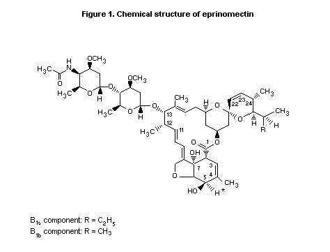
The chemical name of eprinomectin is 4"-deoxy-4"-epiacetylamino-
avermectin B1. It is a semi-synthetic member of the avermectin
family of macrocyclic lactones and consists of a mixture of two
homologous components, B1a (not less than 90%) and B1b (not more
than 10%), which differ by a single methylene group at C26. The
structure is shown in Figure 1. The purity of the compound used in the
studies of toxicity was determined to be 95.1-99.6% by
high-performnace liquid chromatography (HPLC).
 Eprinomectin is active in animals against internal and external
parasites. Its precise mode of action, in common with other
avermectins, is unknown, despite many years of investigation of a
variety of compounds in this class. The effect of avermectins,
including eprinomectin, is mediated via a specific, high-affinity
receptor present in the target organisms. The physiological response
to avermectin binding is increased membrane permeability to chloride
ions, which is independent of gamma-aminobutyric acid (GABA)-mediated
chloride channels. Although avermectins interact with the GABA-gated
channels, they do so only at very high concentrations, i.e. about
three orders of magnitude greater than that necessary to activate the
high-affinity receptor. Therefore, the action of the avermectins at
the GABA-gated chloride ion channels is probably not involved in their
nematocidal and insecticidal activity at therapeutic doses. Activation
of the specific avermectin high-affinity receptor ultimately results
in paralysis and death of the target organism (Turner & Schaeffer,
1989). The fact that much higher concentrations of these compounds are
needed in mammals than in nematodes to affect neurological function
may be due to lack of a specific, high-affinity site associated with
neuronal function or to the relatively poor penetration of these
high-compounds into the central nervous system (Lankas & Gordon,
1989).
2. BIOLOGICAL DATA
2.1 Biochemical aspects
2.1.1 Absorption, distribution, and excretion
Rats
[5-3H]Eprinomectin (specific activity, 7400 dpm/µg) was
administered orally by gavage in 0.5% aqueous methylcellulose to
Crl:CD (SD) BR VAF rats at a dose of 6 mg/kg bw per day for one week.
Three rats of each sex were sacrificed 7 h and one, two, and five days
after the final dose. Urine and faeces were collected immediately
before treatment and daily until sacrifice. After sacrifice, samples
of blood, liver, kidneys, abdominal and/or back fat tissue (females)
and/or testicular fat pad (males), hind leg muscles, and
gastrointestinal tract (including contents) were collected. The
radiolabel in each sample was determined by scintillation
spectrometry. The study was certified for compliance with GLP and
quality assurance.
During treatment and the five days thereafter, 90% of the
administered dose was excreted in the faeces and less than 1% in the
urine. The route and rate of excretion were independent of sex. At 7 h
after treatment, the highest total residue concentrations were found
in the gastrointestinal tract (55.6 mg/kg eprinomectin equivalents),
followed by liver (10.7 mg/kg), fat (8.6 mg/kg), kidney (7.6 mg/kg),
and muscle (2.2 mg/kg). Significantly lower concentrations were found
in plasma (0.89 mg/kg) and erythrocytes (0.31 mg/kg). Similar patterns
of distribution were seen at later times. By five days after
treatment, the total residue concentration had declined to
< 0.1 mg/kg in all samples. The depletion pattern was comparable in
male and female rats (Halley et al., 1995).
Cattle
Angus and Hereford beef cattle received single topical
applications of [5-3H]-eprinomectin (as the commercial formulation
Eprinex Pour-On; specific activity, 0.061 mCi/mg or 135 dpm/ng) at a
dose of 0.5 mg/kg bw. Three cattle of each sex were slaughtered 7, 14,
21, and 28 days after treatment. Blood samples were collected from all
animals before treatment and at several times after treatment. Urine
and faeces were collected several times only from cattle slaughtered
at 28 days. After sacrifice, samples of liver, kidney, hindquarter
muscle, muscle beneath the application site, and perirenal fat were
collected; samples of hide at the site of applications were collected
only from those killed at 28 days. The radioactivity in each sample
was determined by scintillation spectrometry; the tissue and plasma
samples were also analysed for eprinomectin B1a by reverse-phase
HPLC. The study was certified for compliance with GLP and quality
assurance.
Eprinomectin was slowly absorbed, as evidenced by a slow rise and
a broad plateau in plasma concentrations over two weeks rather than a
sharp peak. In plasma, the highest total residue concentrations were
in the range 4.4-21.1 ng/ml eprinomectin equivalents and the highest
concentrations of B1a in the range 7.3-20 ng/ml. Only a small
portion of the applied dose was found in the urine (0.35%), and
excretion was mostly in the faeces (14% of the dose after 28 days).
Analysis of the hide samples revealed that 54% of the initially
applied dose remained. By seven days after treatment, the highest
concentrations of total residue were found in liver (980 µg/kg
eprinomectin equivalents), followed by kidney (180 µg/kg), fat
(34 µg/kg), and muscle beneath the application site (24 µg/kg); the
lowest concentrations were found in hindquarter muscle (8 µg/kg). At
later times, the total residue concentrations declined but the
relative concentrations remained the same. By 28 days after treatment,
the total residue concentrations had declined to 185 µg/kg in liver,
30 µg/kg in kidney, 5 µg/kg in fat, 22 µg/kg in muscle beneath the
application site, and 2 µg/kg in hindquarter muscle. The depletion
half-lives for total residues in the different tissues were 7.88.6
days, but that in muscle beneath the application site was 36.1 days;
however, the last value is probably unreliable owing to large
interanimal variation and poor regression fit. In all tissues, the
B1a concentrations accounted for more than 80% of the total
radioactive residues. Depletion of B1a followed the same order as
that of total residues at all times, the depletion half-lives varying
from 7.5-9.6 days in liver, kidney, fat, and muscle to 29.4 days in
muscle beneath the application site. These results indicate that B1a
is depleted in parallel with the total residues in all tissues on days
7-28. The depletion pattern was comparable in male and female cattle
(Green-Erwin et al., 1994).
Holstein dairy cattle were given each of the following four
treatments, with a period of 14 days between treatments: single
intravenous doses of 25, 50, and 100 µg/kg bw eprinomectin in glycerol
formal-propylene glycol and a single topical dose of 0.5 mg/kg bw
eprinomectin in the commercial formulation along the back. Blood
samples were collected from the jugular vein at several times after
each treatment, and the plasma was assayed for eprinomectin by HPLC
with fluorescence detection. The study was certified for compliance
with quality assurance. After intravenous treatment, plasma clearance
was independent of dose, indicating that the concentrations increased
proportionally to dose. The volume of distribution decreased with
increasing dose, corresponding to a decrease in mean residence time.
After topical treatment, maximum plasma concentrations of 17-32 ng/ml
(mean, 21 ng/ml) were reached after 2-5 days (mean, 3.5 days). The
mean residence time was 165 h. The bioavailability was only 29%. Most
of the absorption occurred within 7-10 days after treatment, following
an initial lag of 24 h, but continued for 17-21 days after treatment
(Faidley, 1995).
2.1.2 Biotransformation
Rats
In the study of Halley et al. (1995), described above,
metabolites were identified in all tissue, plasma, and faecal samples
by reverse-phase HPLC with mass spectroscopic analysis. The parent
drug, comprised of B1a and B1b, was the major residue in all
tissues and plasma at 7 h (89-94% in males, 75-93% in females), and in
faeces after one day (87% in males, 82% in females). At these times,
N-deacetylated B1awas the major metabolite in all samples (tissues
and plasma: 0.6-5.2% in males, 2.3-20% in females; faeces: 1.2% in
males, 5.8% in females) and was usually the main residue at later
times (26 and 73% in liver and kidney at two days and 20 and 63% in
faeces at five days in males and females, respectively). Other minor
metabolites, each representing < 7% of the total radiolabel, were
also present in the samples. Three were identified as the 24a-
hydroxymethyl, 24a-hydroxy, and 26a-hydroxymethyl metabolites of
B1a. These results indicate that the primary route of metabolism of
eprinomectin in rats is via N-deacetylation and that eprinomectin is
metabolized more extensively in female than in male rats.
Cattle
The nature of the residues in tissues, plasma, and faeces of
cattle after pour-on administration of [5-3H]eprinomectin at 0.5
mg/kg bw was investigated by reverse-phase HPLC. The study was
certified for compliance with GLP and quality assurance. Eprinomectin
is not extensively metabolized in cattle, as the parent drug was the
main residue at all slaughter times in all tissues (90-95%), plasma
(95%), and faeces (86%). The parent drug contained 78-87% B1a and
7.2-9.3% B1b. N-Deacetylated B1a was a minor metabolite in these
samples (< 1.3%, except for hindquarter muscle which contained 3.9%).
Other minor metabolites present in the samples represented 0.1-2.4% of
the total radiolabel in tissues and plasma and 0.5-7.4% of that in
faeces. The metabolite profile was qualitatively and quantitatively
independent of sex, slaughter time, and tissue type. Thus, shortly
after drug administration, the metabolism of eprinomectin in cattle is
very similar to that in rats, the parent compound representing most
the residue. In rats, however, the amount of the N-deacetylated
metabolite increases relative to total residue at later times, while
in cattle the concentration of this metabolite to total residue
remains relatively constant over time. The profile of other minor
metabolites is qualitatively similar in the two species (Venkataraman
& Narasimhan, 1995).
2.2 Toxicological studies
2.2.1 Acute toxicity
The acute oral and intraperitoneal toxicity of eprinomectin was
studied in groups of three female Crl:CD-1 (ICR) BR mice and female
Crl:CD (SD) BR rats given 9.8, 20, 39, or 78 mg/kg bw. The oral doses
were given by gastric intubation and the intraperitoneal doses by
injection through the ventral abdominal wall. In both cases, the
vehicle was 0.5% aqueous methylcellulose. The study was certified for
compliance with GLP and quality assurance. The approximate value for
the oral LD50 was 70 mg/kg bw for mice and 55 mg/kg bw for rats; in
both species, the approximate intraperitoneal LD50 value was 35
mg/kg bw. The toxic symptoms observed were ataxia, tremors, loss of
righting reflex, ptosis, and bradypnoea. The surviving animals
recovered within four to five days (Bagdon & McAfee, 1990).
2.2.2 Short-term toxicity
Rats
In a 23-day exploratory toxicity study, groups of five male and
five female Crl:CD (SD) BR albino rats received eprinomectin in the
diet at doses of 0, 0.5, 2.5, 5, or 10 mg/kg bw per day. The low dose
was increased to 20 mg/kg bw per day from day 15 onwards. No
treatment-related effects were seen on mortality or clinical signs.
Decreases in weight gain and feed efficiency were observed in female
rats at 20 mg/kg bw per day but not in females at lower doses. No
adverse effects were observed in male rats (Kloss & Morrissey, 1990a).
In a second exploratory study, groups of five male and five
female Crl:CD (SD) BR albino rats received eprinomectin in the diet at
doses of 0, 20, 40, or 60 mg/kg bw per day for 26 days. Owing to
severe clinical signs (ataxia, tail and whole-body tremors, a hunched,
unthrifty appearance, and piloerection), body-weight loss, and
decreased food consumption, the groups at 40 and 60 mg/kg bw per day
were terminated after one week of treatment, and a new group receiving
30 mg/kg bw per day was started. In this group, clinical signs similar
to but milder than those in animals at the two higher doses were
observed, in addition to decreases in body-weight gain and food
consumption. At 20 mg/kg bw per day, male rats were unaffected, but
female rats had moderate reductions in body-weight gain and food
consumption (Kloss & Morrissey, 1990b).
Groups of 20 male and 20 female Crl:CD (SD) BR albino rats
received eprinomectin in the diet for 90 days at nominal doses of 0,
1, 5, or 30 mg/kg bw per day; however, owing to low food consumption
by animals at the highest dose, the actual intake was 25 mg/kg bw per
day. As this dose resulted in a high incidence of whole-body tremors
and large decreases in body-weight gain, the dose was lowered to 20
mg/kg bw per day in week 4 for females and in week 5 for males. The
actual mean intakes throughout study were 0, 1, 5 and 22 mg/kg bw per
day. The study was of conventional design, with GLP and quality
assurance certification.
Two rats at the high dose died under anaesthesia, and one rat
died of trauma due to a maxillofacial fracture. Aside from tremors, no
treatment-related clinical or ophthalmoscopic signs were noted in rats
at 30/20 mg/kg bw per day. Treatment-related effects in males and
females at the high dose included decreased food consumption and
body-weight gain and increased blood urea nitrogen without a
corresponding increase in creatinine. Females also showed decreased
mean lymphocyte values. Additionally, slight increases in urine
specific gravity (males and females), haematocrit and erythrocyte
count (males), serum protein and albumin (females), and slight
decreases in urine volume (males and females) suggest
haemoconcentration at the high dose, probably as a secondary effect of
the decreased food and water intake. Females at the high dose showed
increased absolute and relative (to body and brain weight) weights of
the liver, uterus, pituitary, and adrenal and decreased ovarian,
spleen, and thymic weights. Males at this dose had increased adrenal
weights and reduced weights of thymus, spleen, and prostate.
Histopathological examination showed arrest of normal ovarian
follicular maturation in 15 of 20 females at the high dose, and the
uteri of four animals showed endometrial squamous metaplasia. These
effects are indicative of oestrogen-progesterone imbalance, which was
also manifested in decreased remodelling of the femora (primary
spongiosa) in 12 of 20 females at the high dose. No remarkable changes
were seen in the brain or spinal cord, but slight degeneration of the
sciatic nerves was noted in three males and three females at the high
dose. There were no other morphological changes related to treatment.
The NOEL was 5 mg/kg bw per day (Kloss et al., 1990a).
Dogs
In a six-week exploratory study, groups of two male and two
female beagle dogs received eprinomectin at doses of 0, 0.5, 1, 2, or
4 mg/kg bw per day. For the first 13 days of the study, eprinomectin
was given in the diet; however, because of its unpalatability in
milled dog food, resulting in reduced food consumption and body-weight
loss in the groups at the two highest doses, it was given by gavage in
0.5% aqueous methylcellulose from day 14 onwards. Treatment with the
highest dose was discontinued after the first gavage dose because of
severe clinical effects, consisting of mydriasis, salivation, ataxia,
decreased activity, and the death of one animal. Mydriasis was
occasionally seen in dogs at 2 mg/kg bw per day, and these animals
also had decreased food intake and body weight. No drug-related
changes were seen in dogs at the lower doses (Kloss & Bagdon, 1990).
Groups of four male and four female beagle dogs received
eprinomectin by gavage for 90 days at nominal doses of 0, 0.5, 1, or 3
mg/kg bw per day in 0.5% aqueous methylcellulose. The high dose was
lowered to 2 mg/kg bw per day from week 2 onwards because of toxicity.
The actual doses administered, on the basis of analytical results,
were about 80% of the nominal, resulting in 0, 0.4, 0.8, or
2.4/1.6 mg/kg bw per day. The study had a conventional design, with
GLP and quality assurance certification. During week 1 of treatment,
the dose of 2.4 mg/kg bw per day induced the death of two males,
mydriasis, emesis, ataxia, salivation, lateral recumbency, and
body-weight loss. Once this dose was lowered to 1.6 mg/kg bw per day,
no treatment-related clinical signs or mortality were observed, but
decreased food consumption and body-weight gain were still seen. The
body-weight gain and food consumption of animals at the intermediate
and low doses were comparable to those of controls. No
treatment-related effects were seen on ophthalmoscopic,
electrocardio-graphic, haematological, blood biochemical, or urinary
parameters or on organ weights or gross appearance. Apart from slight
axonal degeneration in the sciatic nerves of two females at the high
dose, no treatment-related microscopic changes were seen in any
tissue, including brain and spinal cord. The NOEL was 0.8 mg/kg bw per
day on the basis of axonal degeneration in the sciatic nerve and
body-weight loss (Kloss et al., 1990b).
In a one-year study, groups of four male and four female beagle
dogs received eprinomectin by gavage at doses of 0, 0.5, 1, or 2 mg/kg
bw per day in 0.5% aqueous methylcellulose. The study had a
conventional design, with GLP and quality assurance certification. The
only clinical sign attributable to treatment was mydriasis in dogs at
the high dose. One animal at this dose became less active, with
salivation and ataxia progressing to lateral recumbency, and was
therefore necropsied in week 13. This animal also had decreased food
intake and weight loss, while no changes in food consumption or body
weight were seen in any other treated dog. Ophthalmoscopic and
electrocardiographic examinations, haematology, blood biochemistry,
urinalysis, and measurement of organ weights indicated no drug-related
changes. Gross findings were limited to pin-point dark-brown or black
foci in the mucosa of the neck of the gall-bladder, which was found
microscopically to be related to inspissated bile, with no changes in
the histology of the gall-bladder or liver. This finding was observed
in 1/8, 1/8, 1/8, and 3/8 animals at 0, 0.5, 1, and 2 mg/kg bw per
day, respectively, and was considered not to be related to treatment.
Histopathological examination showed very slight focal degeneration of
one to three neurons per dog in the pons area and/or the cerebellar
nuclei in three of eight dogs at the high dose. This degenerative
change was characterized by neuronal enlargement resulting from
increased eosinophilic, vacuolated cytoplasm with nuclear
displacement, and was not seen in other treated dogs or controls. No
other remarkable histopathological findings were seen in other
tissues, including spinal cord and sciatic nerves. The NOEL was 1
mg/kg bw per day on the basis of mydriasis and focal neuronal
degeneration in the brain (Kloss et al., 1994).
2.2.3 Genotoxicity
The results of studies of the genotoxicity of eprinomectin are
summarized in Table 1. The studies were of conventional design, with
GLP and quality assurance certification.
2.2.4 Reproductive toxicity
(i) Multigeneration reproductive toxicity
Rats
In a range-finding study of reproductive toxicity, groups of 15
female Crl:CD (SD) BR rats received eprinomectin at dietary
concentrations of 0, 7, 36, or 180 mg/kg feed per day for 16 days
before cohabitation, during cohabitation, and from day 0 of gestation
through day 21 of lactation. When cohabitation lasted more than one
night, eprinomectin was administered once daily by oral gavage in 0.5%
aqueous methylcellulose; this occurred only in rats at the low and
intermediate doses. On the basis of food intake, the overall mean
intake of eprinomectin was 0, 0.7, 3.3, and 13 mg/kg bw per day,
respectively. Females were mated with untreated males and were allowed
to deliver naturally. Dams and pups were killed within two days of day
21 of lactation. The study was certified for compliance with GLP and
quality assurance.
Dams showed no treatment-related deaths, abortions, or physical
signs, and no effects were seen on length of gestation, the percent of
females with live pups, or the percent of live pups at birth. Females
at the intermediate dose had increased body-weight gain during days
020 of lactation because of failure to lose weight on days 812 of
lactation, as is normal. In comparison with controls, dams at the high
dose had decreased body-weight gain throughout treatment and slightly
decreased food consumption on gestation days 0-8 and lactation days
04. These animals were killed before lactation day 8 because of
excessive pup mortality. They also showed significantly decreased
fecundity indexes, number of implants per female, percent
postimplantation survival, and number of live pups per litter.
External examination of the pups revealed no treatment-related
effects, but increased pup mortality was observed at the highest dose,
particularly during lactation days 4-7. The remaining pups, which all
had tremors, were therefore killed on lactation day 8. At the
intermediate dose, toxicity in pups was evidenced by decreased body
weight and fine tremors during the middle and end of lactation
(Cukierski, 1990a).
Table 1. Results of assays for genotoxity with eprinomectin
End-point Test object Concentration Result Reference
In vitro
Reverse S. typhimurium TA97a, 100-10 000 Negativea Sina (1990,
mutation TA98, TA100, TA 1535 µg/plate 1994)
E. coli WP2, WP 2 uvrA,
WP2 urA pKM101
Gene mutation V-79 Chinese hamster 1-40 µmol/ Negativeb DeLuca (1991)
lung cells (hprt locus) plate (-S9)
10-40 µmol/
plate (+S9)
Cytogenetic Chinese hamster ovary 8-12 µmol/ Negativeb Galloway
alterations cells plate (-S() (1990)
5-7 µmol/
plate (+S9)
DNA damage Primary rate hepatocytes 10-51 µmol/ Negative Storer (1990)
plate
In vivo
Micronucleus Mouse bone marrow 10-40 mg/kg Negativec Galloway
formation bw, once by (1994)
oral gavage
a With and without rate liver S9 fraction; precipitation on all plates at 10 000 µg/plate
b Dose-related cytotoxicity with and without rate liver S9 fraction
c At all doses and all times, the ratio of polychromatic to normochromatic erythrocytes did not
deviate from that in controls; however, clinical signs of toxicity (including decreased
activity, ataxia, and tremors) were observed at the highest dose.
In a two-generation study of reproductive toxicity, groups of 32
male and 32 female Crl:CD (SD) BR VAF/Plus rats received diets
containing eprinomectin at 0, 6, 18, or 54 mg/kg feed. Treatment
started 10 (males) or two (females) weeks before mating and was
continued until all litters had been weaned. An F1 generation of 28
animals of each sex per dose was selected and treated directly from
four weeks of age. These animals were mated at 16 weeks of age to
produce the F2a generation. After being allowed to rear their
litters, the F1 animals were remated at 27 weeks of age to produce
the F2b generation. In order to investigate body tremors in the
offspring, the dietary concentrations of eprinomectin for the F1
animals were reduced to 50% of their initial values during lactation
of the F2b offspring. A contingent of 24 F2b animals of each sex
per dose (except for those at 54 mg/kg, owing to inadequate numbers)
was treated directly during weeks 4-7 of age, after which they were
killed. The brain, spinal cord, and sciatic nerves of F0 and F1
adults killed at about 27 and 38 weeks of age, respectively, and of
F2b pups killed on day 21 post partum were examined
histologically. The study was of conventional design, with GLP and
quality assurance certification.
F0 animals at all doses had slightly increased food consumption
only during the first two weeks of treatment, resulting in slightly
increased body weights. As this effect was transient and small, it is
not considered toxicologically significant. Treatment at 6 mg/kg feed
had no adverse effects on parents or their offspring. Treatment at 18
mg/kg feed resulted only in body tremors in F2a pups in four of 26
litters after day 8 of lactation. Treatment at 54 mg/kg feed had
adverse effects on the dams, their reproduction, and their litters. No
treatment-related deaths or physical signs occurred among the parental
animals. The F1 animals had lowered body weights at week 4,
reflecting their impaired growth during the pre-weaning period. During
the first weeks of treatment, the food consumption and body weights of
F1 animals were decreased, but these differences tended to be
abolished or even reversed in later phases of the study. Although
within each treated group, food consumption during lactation was
increased over that during gestation, the food consumption of F0 and
F1 (first mate) females was reduced during the first two weeks of
lactation in comparison with controls; a similar effect, although less
marked, was observed after the second mate of the F1 animals at the
reduced dose of 27 mg/kg feed. Sexual maturation was delayed in F1
animals, consistent with their delayed physical development. After the
first mating of the F1 generation the pregnancy rate was slightly
reduced, and at the second mating of these animals there was marked
impairment of mating performance and a 50% reduction in pregnancy
rate, resulting in a reduction in the number of females producing live
litters. Litter sizes were not affected by treatment. Signs of
toxicity in F1 and F2a pups were markedly increased mortality
after day 8 post partum, decreased litter and mean pup weights from
day 8 post partum through to weaning, and body tremors in all pups
in all litters. In F2b pups, no body tremors were observed at any
dose when the dietary concentrations were reduced to 0, 3, 9, or 27
mg/kg feed, and the pup losses were not different from those of
controls; however, at 27 mg/kg feed, the litter and mean pup weights
were decreased, but to a lesser degree than for F1 and F2a pups.
The NOEL for maternal toxicity was 18 mg/kg feed, equal to 2.5
mg/kg bw per day, on the basis of decreased food intake during the
first two weeks of lactation in F0 and F1 dams. The NOEL for
reproductive toxicity was 18 mg/kg feed, equal to 1.6 mg/kg bw per
day, on the basis of impaired reproductive performance in the F1
animals. On the basis of tremors in F2a pups and decreased body
weights in F2b pups, the NOEL for pup toxicity was 9 mg/kg feed,
equal to 1.3 mg/kg bw per day (Brooker et al., 1992).
In a follow-up study to determine the concentrations of
eprinomectin in maternal plasma and milk, groups of 12 mated female
Crl:CD (SD) BR rats received eprinomectin at dietary concentrations of
0, 6, 54/27, or 54 mg/kg feed from day 15 of gestation through day 21
of lactation. The group at the intermediate dose received 54 mg/kg
feed from day 15 of gestation through parturition but 27 mg/kg feed
from day 0 of lactation through sacrifice to compensate for increased
maternal food consumption during lactation. The actual mean intakes of
eprinomectin during gestation were 0, 0.4, 4.0, and 4.1 mg/kg bw per
day, and those during lactation were 0, 1.2, 4.5, and 6.6 mg/kg bw
per day, respectively. All females were allowed to deliver naturally,
and dams and pups were killed within four days of day 21 of lactation.
The study was certified for compliance with GLP and quality assurance.
No treatment-related deaths, abortions, or physical signs were
seen among the dams, and there were no effects on the length of
gestation or the number of live pups per pregnant female. In
comparison with controls, the body-weight gain of dams at the high
dose was increased during days 15-21 of gestation and days 0-21 of
lactation, and the food consumption of dams at the intermediate and
high doses was decreased during lactation days 8-21. Within the groups
at the intermediate and high doses, however, food consumption was
increased from gestation day 15 through lactation day 4. Eprinomectin
was well absorbed by all rats, sustained concentrations being detected
in milk and maternal plasma during lactation days 7-21 with a direct
doseconcentration relationship: the overall milk:plasma ratio was
approximately 3:1. Treatment with eprinomectin resulted in
dose-dependent toxicity in pups at the intermediate and high doses
starting on or after day 5 of lactation. The signs of toxicity were
decreased body-weight gain and intermittent body tremors in pups at
the intermediate and high doses and increased mortality (mainly on
lactation days 8-14) among pups at the high dose. As these effects
were observed during a period when the only route of exposure was
through milk, they are probably due to postnatal exposure, as
evidenced by the sustained concentrations of eprinomectin in milk and
as further supported by the results reported below (Mattson, 1992).
In a multigeneration study of reproductive toxicity in rats with
the related compound ivermectin at oral doses of 0.05-3.6 mg/kg bw per
day, ivermectin had no effect on mating, fertility, or pregnancy up to
the highest dose tested. Similar neonatal toxicity, characterized by
decreased weight gain and pup mortality during lactation, was,
however, observed in offspring at doses > 0.4 mg/kg bw per day, with
a NOEL of 0.2 mg/kg bw per day. In a cross-fostering study, it was
shown that the neonatal toxicity was not related to exposure in
utero but to postnatal exposure through the milk. The concentrations
of ivermectin (a highly lipophilic compound) in milk were three to
four times those in plasma. These relatively high concentrations of
ivermectin in milk resulted in significantly higher concentrations in
the brain and plasma of nursing offspring, and the period of enhanced
neonatal sensitivity correlated with the increased plasma:brain ratios
of ivermectin, consistent with postnatal formation of the blood-brain
barrier in this species. In other mammalian species, including humans,
the blood-brain barrier is formed prenatally. Therefore, the toxicity
of ivermectin in neonatal rats is probably the result of a combination
of excessive exposure through maternal milk and the increased
permeability of the blood-brain barrier during the early postnatal
period in this species (Lankas & Gordon, 1989; Lankas et al., 1989).
(ii) Developmental toxicity
Rats
In a range-finding study, eprinomectin was administered by gavage
in 0.5% aqueous methylcellulose at doses of 0, 0.5, 1.5, 5, 10, or 15
mg/kg bw per day to groups of 10 mated female Crl:CD (SD) BR rats on
days 6-17 of gestation. Serum biochemical and haematological
examinations were performed on day 14 of gestation. On day 20 of
gestation, the dams were killed and necropsied, and the fetuses were
weighed and examined for external abnormalities. One dam at the high
dose was killed on day 14 of gestation because of severe weight loss;
this animal also had slight tremors, ptosis, decreased activity, and
abnormal posture and had increased erythrocyte count, haemoglobin, and
haematocrit. One rat at the low dose died on day 14 of gestation due
to anaesthesia overdose. There were no abortions. Some of the animals
at the high dose had fine tremors, abnormal posture, and reluctance to
be handled. Maternal body weight gain was significantly increased at 5
and 10 mg/kg bw but significantly decreased at 15 mg/kg bw. The
concentration of urea nitrogen and the activity of alanine
aminotransferase were increased in rats at the two highest doses. No
effects were observed on haematological parameters or on the number of
implants, resorptions, or live or dead fetuses. Fetal body weights
were significantly decreased at 1.5, 5, 10, and 15 mg/kg bw, but there
was no dependence on dose. This effect was not seen in the main study,
with larger groups (see below). External examination of the fetuses
showed no evidence of teratogenicity (Cukierski, 1990b).
In the main study, groups of 25 mated female Crl:CD (SD) BR rats
were treated orally by gavage with eprinomectin in 0.5% aqueous
methylcellulose at doses of 0, 0.5, 1, 3, or 12 mg/kg bw per day on
days 6-17 of gestation. On day 20 of gestation, the dams were killed
and necropsied, and the fetuses were weighed, sexed, and examined for
external, visceral, and skeletal abnormalities. The study was of
conventional design, with GLP and quality assurance certification.
There were no treatment-related physical signs, deaths, abortions, or
gross lesions. Increased weight gain and food consumption were
observed during treatment with the two highest doses, followed by
decreases during days 18-20, resulting in slightly increased total
weight gain on days 6-18 of gestation. There was no evidence of
developmental toxicity or teratogenicity at doses up to 12 mg/kg bw
per day on the basis of postimplantation survival, fetal weight, and
external, visceral, and skeletal examination. The NOEL for maternal
toxicity was 1 mg/kg bw per day, on the basis of changes in body
weight and food consumption. The NOEL for developmental toxicity was
12 mg/kg bw per day, the highest dose tested (Cukierski, 1991).
Rabbits
In a range-finding study, groups of six female New Zealand white
rabbits received eprinomectin in 0.5% aqueous methylcellulose by
gavage at doses of 0, 1.5, 4, 10, or 25 mg/kg bw per day for 14 days.
Owing to excessive weight loss and poor condition, the animals at 10
and 25 mg/kg bw per day were killed on days 8 and 3 of treatment,
respectively. There were no deaths and no effects on body weight at
the lower doses. Dilated pupils and slowed pupillary reflexes were
observed at doses > 4 mg/kg bw per day, and mild tremors and
decreased food consumption were seen at doses > 10 mg/kg bw per
day. At 25 mg/kg bw per day, some animals neither urinated nor
defaecated (Clark, 1990).
In a second range-finding study, eprinomectin was administered by
gavage in 0.5% aqueous methylcellulose at doses of 0, 2, 4, or 8 mg/kg
bw per day to groups of eight inseminated female New Zealand white
rabbits on days 6-18 of gestation. Serum biochemical and
haematological examinations were performed on day 19 of gestation. On
day 28 of gestation, the dams were killed and necropsied, and the
fetuses were weighed and examined for external abnormalities.
Treatment with eprinomectin was associated with mydriasis and slowed
pupillary reflex in all groups, and unresponsive mydriasis was found
in the groups at the two highest doses. On day 12 of gestation, one
rat at the high dose died from an intubation accident, and two others
at this dose were killed on days 19 and 27 of gestation because of
severe weight loss after not eating for one week. Slightly decreased
food consumption and weight gain were also observed in the remaining
rats at the high dose and in those at the intermediate dose. No
effects were found on haematological or blood biochemical parameters
or on the numbers of implants, resorptions, or live or dead fetuses,
or on fetal body weights. External examination of the fetuses revealed
no treatment-related findings (Minsker, 1990).
In the main study, groups of 18 inseminated female New Zealand
white rabbits were treated orally by gavage with eprinomectin in 0.5%
aqueous methylcellulose at doses of 0, 0.5, 2, or 8 mg/kg bw per day
on days 6-18 of gestation. On day 28 of gestation, the dams were
killed and necropsied, and the fetuses were weighed, sexed, and
examined for external, visceral, and skeletal abnormalities. The study
was of conventional design, with GLP and quality assurance
certification. There were no treatment-related deaths, abortions, or
gross lesions. Maternal toxicity was evidenced by slowed pupillary
reflex at the intermediate and high doses and mydriasis non-responsive
to light and a slight decrease in body-weight gain in rabbits at the
high dose. The numbers of implants and live fetuses per pregnant
female were decreased at 2 and 8 mg/kg bw per day (significantly only
at the highest dose), but these findings were considered not to be
treatment-related, because the values were still within the range in
historical controls and the lower values were a consequence of fewer
corpora lutea per female at these doses. Likewise, the apparent
increase in the percent preimplantation loss in animals at the
intermediate and high doses was due to the smaller number of implants
and was considered not to be treatment-related. There was no effect on
live fetal weight, and there was no indication of teratogenicity at
doses up to 8 mg/kg bw per day. The NOEL for maternal toxicity was 0.5
mg/kg bw per day on the basis of slowed pupillary reflex. The NOEL for
developmental toxicity was 8 mg/kg bw per day, the highest dose tested
(Wise, 1991).
In order to re-examine the possible effects of eprinomectin on
embryo and fetal viability, a second study was conducted with larger
groups. Eprinomectin was administered by gavage in 0.5% aqueous
methylcellulose at doses of 0, 1.2, 2, or 8 mg/kg bw per day to groups
of 24 mated female New Zealand white rabbits on days 6-18 of
gestation. After sacrifice of the dams on day 28 of gestation, the
numbers of corpora lutea, implants, resorptions, and live or dead
fetuses were counted. The fetuses were not examined further. The study
was certified for GLP and quality assurance. There were no
treatment-related deaths or abortions. Maternal toxicity was seen only
in rabbits at the high dose, which showed slowed pupillary reflex
and/or mydriasis and decreased body-weight gain during treatment. No
effects were found on embryonic or fetal survival. The NOEL for
maternal toxicity was 2 mg/kg bw per day on the basis of physical
signs and decreased body-weight gain. The NOEL for developmental
toxicity was 8 mg/kg bw per day (Cukierski, 1994).
2.2.5 Special studies on target animals
The safety of the commercial formulation Eprinex Pour-On
(containing eprinomectin in Myglyol 840 and 0.01% butylated
hydroxytoluene) was tested by topical application to calves and
breeding animals. Eight-week-old calves were treated at once, three
times, or five times the recommended dose three times at seven-day
intervals, while 12-month-old calves were treated once at 10 times the
recommended dose. Breeding bulls were treated once at three times the
recommended dose, and breeding cows were treated with at least three
times the recommended dose throughout the reproductive cycle. The
studies were certified for compliance with GLP and quality assurance.
In all studies, eprinomectin was well tolerated and was without
adverse effects (Gogolewski, 1994; Bierschwal, 1995; Bridi, 1995;
Pitt, 1995).
2.2.6 Toxicity of emamectin
The toxicology of emamectin has also been reviewed (Department of
Health and Family Services, 1997). Like eprinomectin, emamectin is an
amino-substituted avermectin; the only difference between the two
compounds is the presence of an epi-methylamino group at the C4
position on the emamectin molecule, rather than an epi-acetylamino
group at that position in the case of eprinomectin. The following
studies of the short-term and long-term toxicity of emamectin were
extracted directly from the review.
Mice
Groups of mice were given emamectin at doses of 0.5, 2.5, or
12.5 mg/kg bw per day in the diet for 547-550 days. The dose of 12.5
mg/kg bw per day was reduced to 7.5 mg/kg bw per day in females during
week 48, to 7.5 mg/kg bw per day in males during week 9, and further
reduced to 5.0 mg/kg bw per day in males during week 31. The mortality
rate was increased in males and females at 12.5/7.5/5.0 mg/kg bw per
day. Tremors and vocalization was seen in three to four male mice
treated with 12.5 mg/kg bw per day between weeks 5 and 8-9, but these
adverse clinical signs abated after the dose was reduced to 7.5 mg/kg
bw per day. Vocalization occurred in female mice treated with 12.5
mg/kg bw per day after week 16, but was not evident after week 34.
Several animals treated with 12.5/7.5/5.0 mg/kg bw per day developed
minor neurological abnormalities, e.g. fine forelimb fasciculations,
after week 14, which persisted until the end of the study. Two males
given 12.5 mg/kg bw per day had sciatic nerve degeneration,
characterized by vacuolation and the presence of myelin balls in the
nerve fibres. The body-weight gain of males and females was reduced
after one to two weeks of treatment with 12.5/7.5/5.0 mg/kg bw per
day. Emamectin showed no carcinogenic potential. The NOEL was 2.5
mg/kg bw per day on the basis of neurological abnormalities and
decreased weight gain in mice receiving higher doses.
Rats
Groups of rats were given emamectin at 0, 0.5, 2.5, or 12.5/8/5
mg/kg bw per day in their diet for 14 weeks. The dose of 12.5 mg/kg bw
per day was reduced to 8 mg/kg bw per day during week 3 and
subsequently to 5 mg/kg bw per day during week 9. During weeks 3-11 of
treatment, nine of 20 males receiving 12.5/8/5 mg/kg bw per day were
killed because of ill health. Genera-lized body tremors were noted in
most animals receiving 12.5/8/5 mg/kg bw per day, but the incidence
decreased as the dose was lowered. During week 7, splaying of the
hindlimbs was seen in a number of males and females receiving 8 mg/kg
bw per day, which was associated with histological lesions in nervous
tissue. Significant reductions in body weight and food consumption
were seen in animals receiving 12.5/8/5 mg/kg bw per day. Decreased
serum glucose concentration and a slight increase in blood urea
nitrogen were seen at all sampling times in males and females
receiving 12.5/8/5 mg/kg bw per day. Decreased urine output and an
increase in urine specific gravity were seen in groups receiving
12.5/8/5 mg/kg bw per day; at the same dose, neuronal cytoplasmic
vacuolation and degeneration were noted. The NOEL was 2.5 mg/kg bw per
day on the basis of neurotoxicity, weight loss, and decreased food
consumption in rats receiving higher doses.
Groups of rats were given emamectin at doses of 0, 0.1, 1.0, 2.5
(males), or 5/2.5 (females) mg/kg bw per day in the diet for 53 weeks.
The dose of 5 mg/kg bw per day in female rats was reduced to 2.5 mg/kg
bw per day in study week 18. No treatment-related deaths were seen.
Generalized body tremors were seen in females treated with 5/2.5 mg/kg
bw per day, starting during week 9 and increasing in frequency up to
week 18; tremors were not seen after week 21 and were not reported in
males at doses < 2.5 mg/kg bw per day. A reduction in body weight
was seen in females given 5 mg/kg bw per day, but after the dose was
reduced to 2.5 mg/kg bw per day the body-weight gain gradually
returned to that of controls, to which it was comparable by week 25.
From week 37, females given 1.0 or 2.5 mg/kg bw per day had a slight
increase in body weight. In general, the weight changes parallelled
the minor decreases and increases in food consumption. The females
given 5 mg/kg bw per day showed decreased forelimb grip strength by
week 14, which decreased in frequency up to and including 24 weeks; no
neurological abnormalities were seen beyond 24 weeks. Neuronal
degeneration of the brain was seen in 19 of 20 females receiving 5/2.5
mg/kg bw per day and 9 of 20 males given 2.5 mg/kg bw per day, and
degeneration of the spinal cord was seen in 2 of 20 females and 4 of
20 males given 5/2.5 and 2.5 mg/kg bw per day, respectively. The NOEL
was 1 mg/kg bw per day on the basis of neurological toxicity in rats
receiving higher doses.
Groups of rats were given emamectin at doses of 0.25, 1, or 5/2.5
mg/kg bw per day in the diet for 105 weeks. The dose of 5 mg/kg bw per
day was reduced to 2.5 mg/kg bw per day in week 6 for males and week
10 for females. Weight gain and food consumption were increased in
females given doses > 1 mg/kg bw per day. Serum triglyceride
concentrations were elevated in animals fed 1 and 5/2.5 mg/kg bw per
day for most of the study, and elevated serum bilirubin concentrations
were seen in the latter half of the study in females fed 1 or 5/2.5
mg/kg bw per day. Males at the highest dose showed reduced weight gain
and food intake in the latter half of the study. Neuronal vacuolation
was seen in the brain and spinal cord of male and female rats given
5/2.5 mg/kg bw per day, and an increased incidence of diffuse
vacuolation of hepatocytes was seen in female rats fed 1 or 5/2.5
mg/kg bw per day. Emamectin had no carcinogenic potential. The NOEL
was 0.25 mg/kg bw per day on the basis of increased weight gain, food
consumption and serum triglyceride and bilirubin concentrations in
rats receiving higher doses.
Dogs
Groups of beagle dogs were given emamectin at doses of 0, 0.5, 1,
or 1.5 mg/kg bw per day for 14 weeks, but the doses were reduced to
0.25, 0.5, and 1 mg/kg bw per day respectively, at the start of week
3. Three animals in the group receiving 1.5/1 mg/kg bw per day were
killed during weeks 3-6 of treatment after showing tremors, mydriasis,
anorexia, and lethargy. Six of eight animals receiving 1.5/1 mg/kg bw
per day had tremors, mostly beginning during week 2 of treatment.
Animals at 1.5 mg/kg bw per day had reduced weight gain and food
consumption, but these parameters returned to normal when the dose was
reduced to 1 mg/kg bw per day. Treatment-related histological changes
were seen in the brain, spinal cord, sciatic and optic nerves, and
skeletal muscle. Neuronal degeneration was seen in the brains of all
animals receiving 1.5/1 mg/kg bw per day and in 50% of animals
receiving 1/0.5 mg/kg bw per day. Scattered neuronal vacuolation was
noted in the spinal cords of all animals treated with 1.5/1 mg/kg bw
per day and of one of eight animals treated with 1/0.5 mg/kg bw per
day. Sciatic and optic nerve lesions consisting of scattered
vacuolation were seen in most animals receiving 1.5/1 mg/kg bw per
day. Very slight to moderate skeletal muscle atrophy was seen in seven
of eight animals receiving 1.5/1 mg/kg bw per day and two of eight
animals receiving 1/0.5 mg/kg bw per day. The NOEL was 0.25 mg/kg bw
per day on the basis of neuronal degeneration and skeletal muscle
atrophy in dogs receiving higher doses.
Groups of dogs were given emamectin at doses of 0, 0.25, 0.5,
0.75, or 1 mg/kg bw per day by gavage for 53 weeks. Owing to evidence
of overt toxicity, in the form of body tremors, mydriasis, decreased
motor activity, and reduced food consumption and body weight, all
animals receiving 1 mg/kg bw per day were killed on day 23 of
treatment. Most of the males at 0.75 mg/kg bw per day developed
tremors, stiffness of gait, mydriasis, and weight loss and were killed
on day 50 of the study. Similar signs to those in males were seen in
females treated with 0.75 mg/kg bw per day, but they were of decreased
severity. One of eight dogs in the group treated with 0.5 mg/kg bw per
day had fine tremors. Those given 1 mg/kg bw per day, particularly the
female animals, showed weight loss associated with decreased food
intake. Three of four males treated with 0.75 mg/kg bw per day had
weight loss and decreased food consumption. Neuronal degeneration in
the central nervous system was reported in males treated with 0.75
mg/kg bw per day and males and females treated with 1 mg/kg bw per
day. Axonal degeneration in the central and peripheral nervous systems
was seen at doses > 0.5 mg/kg bw per day in animals of each sex.
Degeneration of the retinal ganglionic cells and axonal degeneration
of the optic nerve were reported at doses of 0.75 and 1 mg/kg bw per
day. The NOEL was 0.25 mg/kg bw per day on the basis of neurotoxicity
in dogs receiving higher doses.
3. COMMENTS
The Committee considered the results of studies on the
pharmacokinetics, metabolism, acute and short-term toxicity,
genotoxicity, and reproductive toxicity of eprinomectin. All of the
pivotal studies were carried out according to appropriate standards
for study protocol and conduct.
When radiolabelled eprinomectin was administered orally to rats,
the radiolabel was found mainly in the gastrointestinal tract,
followed by liver, fat, and kidney, while lower levels were found in
muscle and blood. Elimination occurred almost exclusively in the
faeces. For up to 24 h after drug administration, the major residue in
tissues, plasma, and faeces was unchanged eprinomectin. After two to
five days, the major residue was N-deacetylated B1a. The primary
route of metabolism of eprinomectin in rats is thus N-deacetylation,
and minor routes are hydroxylation and hydroxymethylation. Metabolism
is more extensive in female than in male rats. In cattle,
radiolabelled eprinomectin was absorbed slowly after topical
administration. The absorbed radiolabel was taken up mainly by the
liver and to a lesser extent by the kidney, fat, and muscle. The
radiolabel disappeared from these tissues with half-lives of 7.88.6
days, except for muscle beneath the application site in which the
half-life was 36 days. Elimination occurred mostly in the faeces. At
all times of slaughter, the main residue in tissues, plasma, and
faeces was unchanged eprinomectin (85%), B1a representing more than
80%. B1a disappeared in parallel with the total residues in all
tissues at all slaughter times, with half-lives of 7.59.6 days in
liver, kidney, fat, and muscle and 29 days in muscle beneath the
application site. The profile of metabolites in cattle was
qualitatively similar to that in rats.
After oral administration of eprinomectin, the approximate LD50
values were 70 mg/kg bw for mice and 55 mg/kg bw for rats.
Eprinomectin is moderately hazardous after acute oral exposure.
In a 90-day study of toxicity, rats received eprinomectin in the
diet at nominal doses of 0, 1, 5, or 30/20 mg/kg bw per day. Male and
female rats at the high dose had tremors, slight degeneration of the
sciatic nerves, decreased body-weight gain, and changes in organ
weights. Females at this dose also had arrest of normal ovarian
follicular maturation, endometrial squamous metaplasia, and decreased
remodelling of the femora (primary spongiosa), indicative of
oestrogenprogesterone imbalance. The NOEL was 5 mg/kg bw per day on
the basis of effects on the central nervous system and other effects.
In a 90-day study of toxicity, dogs received eprinomectin by
gavage at doses of 0, 0.4, 0.8, or 2.4/1.6 mg/kg bw per day. The
highest dose induced mydriasis, emesis, ataxia, salivation, lateral
recumbency, body-weight loss, or death. After reduction of this dose
to 1.6 mg/kg bw per day, decreased food consumption and decreased
body-weight gain were observed in males and females. Females at this
dose had slight axonal degeneration of the sciatic nerves. The NOEL
was 0.8 mg/kg bw per day on the basis of sciatic nerve axonal
degeneration and body-weight loss.
In a one-year study of toxicity, dogs received eprinomectin by
gavage at doses of 0, 0.5, 1, or 2 mg/kg bw per day. Treatment-related
effects were observed only at the highest dose; these included
mydriasis and slight focal neuronal degeneration in the pons and the
cerebellar nuclei of the brain. On the basis of these effects, the
NOEL was 1 mg/kg bw per day.
Eprinomectin has been tested in vitro for its ability to induce
reverse mutations in Salmonella typhimurium and Escherichia coli,
gene mutations in Chinese hamster lung cells, chromosomal aberrations
in Chinese hamster ovary cells, and DNA single-strand breaks in
primary rat hepatocytes. It has been tested in vivo for its ability
to induce micronuclei in mouse bone marrow. The results of all tests
were negative. On the basis of these data, the Committee concluded
that eprinomectin is unlikely to be genotoxic.
Rats were exposed to eprinomectin at dietary concentrations of 0,
6, 18, or 54 mg/kg feed in a two-generation study of reproductive
toxicity. On the basis of decreased food intake by the dams during the
first two weeks of lactation, the NOEL for maternal toxicity was 18
mg/kg feed, equal to 2.5 mg/kg bw per day. The NOEL for reproductive
toxicity was 18 mg/kg feed, equal to 1.6 mg/kg bw per day, on the
basis of delayed sexual maturation and a reduced pregnancy rate in
first-generation animals. Toxicity to pups was the most sensitive
indicator of the effects of eprinomectin, which consisted of decreased
weights, body tremors, and increased mortality at 54 mg/kg feed in the
first-generation pups and in the second-generation pups of the first
mating. The second-generation pups also had body tremors when treated
at 18 mg/kg feed. No body tremors or deaths occurred in the second-
generation pups of the second mating at any dose when the dietary
levels were reduced to 0, 3, 9, or 27 mg/kg feed, but decreased pup
weights were still seen at 27 mg/kg feed. The NOEL for pup toxicity
was 9 mg/kg feed, equal to 1.3 mg/kg bw per day. Furthermore, no
histopathological changes were observed in the brain, spinal cord, or
sciatic nerves of animals treated up to 38 weeks of age. A follow-up
study suggested that the toxicity to pups is probably due to postnatal
exposure through maternal milk, as there were high, sustained
concentrations of eprinomectin in milk. This conclusion was further
supported by the results of a cross-fostering study with ivermectin, a
closely-related compound, which was reviewed by the Committee at its
thirty-sixth meeting (Annex 1, reference 91).
In studies of developmental toxicity in rats and rabbits,
eprinomectin caused maternal toxicity, evident in rat dams as changes
in body-weight gain and food consumption at oral doses of 3 and 12
mg/kg bw per day, with a NOEL of 1 mg/kg bw per day. Rabbit dams had
slowed pupillary reflexes, mydriasis, and decreased body-weight gain
at oral doses of 2 and 8 mg/kg bw per day, giving a NOEL of 1.2 mg/kg
bw per day. Eprinomectin did not cause embryotoxicity, fetotoxicity,
or teratogenicity in either species at oral doses up to 12 mg/kg bw
per day in rats and 8 mg/kg bw per day in rabbits.
No long-term studies were available on eprinomectin; however, the
long-term toxicity of emamectin, another amino-substituted avermectin
structurally very similar to eprinomectin, has been reported. The
Committee noted that dogs are the most sensitive species to both
emamectin and eprinomectin and that the toxicological end-point for
both compounds is neurodegeneration. It further noted that the
neurotoxic effects of both compounds did not progress with prolonged
treatment, resulting in the same NOELs in 90-day and one-year studies
in dogs. On the basis of this information, the Committee concluded
that it was unnecessary to request long-term studies of the toxicity
of eprinomectin.
Because the chemical structure of eprinomectin contains no
structural alerts, and the structurally closely related avermectins,
emamectin and abamectin, are not carcinogenic in mice or rats, the
Committee concluded that eprinomectin is unlikely to be carcinogenic.
This conclusion was supported by the negative findings in studies of
genotoxicity with eprinomectin in vitro and in vivo.
4. EVALUATION
The Committee considered that the most relevant effect for
evaluating the safety of residues of eprinomectin is the effect on the
mammalian nervous system. An ADI of 0-10 µg/kg bw was established on
the basis of the NOEL of 1 mg/kg bw per day for mydriasis and focal
neuronal degeneration in the brain in the one-year study in dogs and a
safety factor of 100.5.
5. REFERENCES
Bagdon, W.J. & McAfee, J.L. (1990) L-653,648: Acute toxicity studies
in mice and rats. Unpublished report (studies no. TT #90-2512,
TT #90-2513, TT #90-2526, and TT #90-2527) from Merck Sharp & Dohme
Research Laboratories, West Point, Pennsylvania, USA. Submitted to WHO
by MSD Sharp & Dohme GmbH, Haar, Germany.
Bierschwal, C.J. (1995) MK-397, cattle, safety, toxicity,
reproduction, breeding bulls. Unpublished report (trial no. ASR 14148)
from Merck & Co., Inc., Fulton, Missouri, USA. Submitted to WHO by MSD
Sharp & Dohme GmbH, Haar, Germany.
Bridi, A.A. (1995) MK-397, cattle, safety, toxicity, reproduction,
breeding cows. Unpublished report (trial no. 13639) from MSDRL
Uruguaiana Veterinary Research Center, Uruguaiana, RS, Brazil.
Submitted to WHO by MSD Sharp & Dohme GmbH, Haar, Germany.
Brooker, A.J., Myers, D.P. & Parker, C.A. (1992) L-653,648:
Two-generation dietary reproduction study in the rat. Unpublished
report (study no. TT #90-9010) from Huntingdon Research Centre Ltd,
Huntingdon, Cambridgeshire, United Kingdom (report No. MSD 203/911574)
for Merck Research Laboratories, West Point, Pennsylvania, USA.
Submitted to WHO by MSD Sharp & Dohme GmbH, Haar, Germany.
Clark, R.L. (1990) L-653,648 Oral range-finding study in non-pregnant
rabbits. Unpublished report (study no. TT #90-719-2) from Merck Sharp
& Dohme Research Laboratories, West Point, Pennsylvania, USA.
Submitted to WHO by MSD Sharp & Dohme GmbH, Haar, Germany.
Cukierski, M.A. (1990a) L-653,648: Dietary range-finding study in
female rats. Unpublished report (study no. TT #90-709-0) from Merck
Sharp & Dohme Research Laboratories, West Point, Pennsylvania, USA.
Submitted to WHO by MSD Sharp & Dohme GmbH, Haar, Germany.
Cukierski, M.A. (1990b) L-653,648: Oral range-finding study in
pregnant rats. Unpublished report (study no. TT #90-718-1) from Merck
Sharp & Dohme Research Laboratories, West Point, Pennsylvania, USA.
Submitted to WHO by MSD Sharp & Dohme GmbH, Haar, Germany.
Cukierski, M.A. (1991) L-653,648: Oral developmental toxicity study in
rats. Unpublished report (study no. TT #90-718-0) from Merck Sharp &
Dohme Research Laboratories, West Point, Pennsylvania, USA. Submitted
to WHO by MSD Sharp & Dohme GmbH, Haar, Germany.
Cukierski, M.A. (1994) MK-0397: Oral embryo/fetal viability study in
rabbits. Unpublished report (study no. TT #94-707-0) from Merck
Research Laboratories, West Point, Pennsylvania, USA. Submitted to WHO
by MSD Sharp & Dohme GmbH, Haar, Germany.
DeLuca, J.G. (1991) L-653,648: V-79 mammalian cell mutagenesis assay.
Unpublished report (studies no. TT #91-8502, TT #91-8510, and TT #91-
8503) from Merck Sharp & Dohme Research Laboratories, West Point,
Pennsylvania, USA. Submitted to WHO by MSD Sharp & Dohme GmbH, Haar,
Germany.
Department of Health and Family Services (1997) Emamectin.
Toxicological evaluation report, prepared by the Chemical Products
Assessment Section, Therapeutic Goods Administration, Department of
Health and Family Services, Australia. Submitted to WHO by Department
of Health and Family Services, Australia.
Faidley, T. (1995) MK0397, topical, cattle, safety, metabolism,
bioavailability, pharmacokinetics. Unpublished report (trial no. ASR
14640) from Merck Research Laboratories, Somerville, New Jersey, USA.
Submitted to WHO by MSD Sharp & Dohme GmbH, Haar, Germany.
Galloway, S.M. (1990) L-653,648: Assay for chromosomal aberrations in
vitro in Chinese hamster ovary cells. Unpublished report (studies no.
TT #90-8611 and TT #90-8614) from Merck Sharp & Dohme Research
Laboratories, West Point, Pennsylvania, USA. Submitted to WHO by MSD
Sharp & Dohme GmbH, Haar, Germany.
Galloway, S.M. (1994) MK-0397: Assay for micronucleus induction in
mouse bone marrow. Unpublished report (study no. TT #93-8719) from
Merck Research Laboratories, West Point, Pennsylvania, USA. Submitted
to WHO by MSD Sharp & Dohme GmbH, Haar, Germany.
Gogolewski, R.P. (1994) MK397, cattle, safety, tolerance, reactions.
Unpublished report (trial no. 14291) from MSD Veterinary Research &
Development Laboratory, Ingleburn, NSW, Australia. Submitted to WHO by
MSD Sharp & Dohme GmbH, Haar, Germany.
Green-Erwin, M., Venkataraman, K. & Narasimhan, N.I. (1994) Depletion
of radioresidues in tissues of cattle dosed topically with a single
dose of radiolabeled MK-0397 (trial CA-368). Unpublished report (study
no. 93767) from Merck Research Laboratories, Rahway, New Jersey, USA.
Submitted to WHO by MSD Sharp & Dohme GmbH, Haar, Germany.
Halley, B.A., Andrew, N.W., Green-Erwin, M.L. & Zeng, Z. (1995) The
distribution, excretion and metabolism of MK-0397 (L-653,648) in rats
(ADMES-1). Unpublished report (study no. 93587) from Merck Research
Laboratories, Rahway, New Jersey, USA. Submitted to WHO by MSD Sharp &
Dohme GmbH, Haar, Germany.
Kloss, M.W. & Bagdon, W.J. (1990) L-653,648: Exploratory six-week oral
toxicity study in dogs. Unpublished report (study no. TT #89-118-0)
from Merck Sharp & Dohme Research Laboratories, West Point,
Pennsylvania, USA. Submitted to WHO by MSD Sharp & Dohme GmbH, Haar,
Germany.
Kloss, M.W., Bagdon, W.J. & Gordon, L.R. (1994) L-653,648:
Fifty-three-week oral toxicity study in dogs. Unpublished report
(study no. TT #92-116-0) from Merck Research Laboratories, West Point,
Pennsylvania, USA. Submitted to WHO by MSD Sharp & Dohme GmbH, Haar,
Germany.
Kloss, M.W., Coleman, J.B. & Allen, H.L. (1990a) L-653,648:
Fourteen-week oral toxicity study in rats. Unpublished report (study
no. TT #90-037-0) from Merck Sharp & Dohme Research Laboratories, West
Point, Pennsylvania, USA. Submitted to WHO by MSD Sharp & Dohme GmbH,
Haar, Germany.
Kloss, M.W., Coleman, J.B. & Ching, S.V. (1990b) L-653,648:
Fourteen-week oral toxicity study in dogs. Unpublished report (study
no. TT #89-141-0) from Merck Sharp & Dohme Research Laboratories, West
Point, Pennsylvania, USA. Submitted to WHO by MSD Sharp & Dohme GmbH,
Haar, Germany.
Kloss, M.W. & Morrissey, R.E. (1990a) L-653,648: Exploratory four-week
oral toxicity study in rats. Unpublished report (study no.
TT #89-119-0) from Merck Sharp & Dohme Research Laboratories, West
Point, Pennsylvania, USA. Submitted to WHO by MSD Sharp & Dohme GmbH,
Haar, Germany.
Kloss, M.W. & Morrissey, R.E. (1990b) L-653,648: Exploratory 4-week
oral toxicity study in rats. Unpublished report (study no.
TT #90-022-0,-1) from Merck Sharp & Dohme Research Laboratories, West
Point, Pennsylvania, USA. Submitted to WHO by MSD Sharp & Dohme GmbH,
Haar, Germany.
Lankas, G.R. & Gordon, L.R. (1989) Toxicology. In: Campbell, W.C.,
ed., Ivermectin and Abamectin, New York, Springer-Verlag, pp.
89-112.
Lankas, G.R., Minsker, D.H. & Robertson, R.T. (1989) Effects of
ivermectin on reproduction and neonatal toxicity in rats. Food
Chem. Toxicol., 27, 523-529.
Mattson, B.A. (1992) L-653,648: Secretion in rat milk study.
Unpublished report (study no. TT #91-26-0) from Merck Research
Laboratories, West Point, Pennsylvania, USA. Submitted to WHO by MSD
Sharp & Dohme GmbH, Haar, Germany.
Minsker, D.H. (1990) L-653,648: Oral range-finding study in pregnant
rabbits. Unpublished report (study no. TT #90-719-1) from Merck Sharp
& Dohme Research Laboratories, West Point, Pennsylvania, USA.
Submitted to WHO by MSD Sharp & Dohme GmbH, Haar, Germany.
Pitt, S.R. (1995) MK 397, cattle, safety, toxicity, reactions.
Unpublished report (trial no. ASR 14578) from MSDRL Veterinary
Laboratory, Hertford, Hertfordshire, United Kingdom. Submitted to WHO
by MSD Sharp & Dohme GmbH, Haar, Germany.
Sina, J.F. (1990) L-653,648: Microbial mutagenesis assay. Unpublished
report (study no. TT #90-8004) from Merck Sharp & Dohme Research
Laboratories, West Point, Pennsylvania, USA. Submitted to WHO by MSD
Sharp & Dohme GmbH, Haar, Germany.
Sina, J.F. (1994) L-653,648: Microbial mutagenesis; Cytotoxicity/
range-finding assay. Unpublished report (study no. TT #89-8059) from
Merck Research Laboratories, West Point, Pennsylvania, USA. Submitted
to WHO by MSD Sharp & Dohme GmbH, Haar, Germany.
Storer, R.D. (1990) L-653,648: In vitro alkaline elution/rat
hepatocyte assay. Unpublished report (studies no. TT #90-8305,
TT #90-8309, and TT #90-8314) from Merck Sharp & Dohme Research
Laboratories, West Point, Pennsylvania, USA. Submitted to WHO by MSD
Sharp & Dohme GmbH, Haar, Germany.
Turner, M. & Schaeffer, J. (1989) Mode of action of ivermectin. In:
Campbell, W.C., ed., Ivermectin and Abamectin, New York,
Springer-Verlag, pp. 73-88.
Venkataraman, K. & Narasimhan, N.I. (1995) Metabolism of
[3H]-MK-0397 in cattle following a topical application (ADMES-3).
Unpublished report (study no. 93993) from Merck Research Laboratories,
Rahway, New Jersey, USA. Submitted to WHO by MSD Sharp & Dohme GmbH,
Haar, Germany.
Wise, L.D. (1991) L-653,648: Oral developmental toxicity study in
rabbits. Unpublished report (study no. TT #90-719-0) from Merck Sharp
& Dohme Research Laboratories, West Point, Pennsylvania, USA.
Submitted to WHO by MSD Sharp & Dohme GmbH, Haar, Germany.
Eprinomectin is active in animals against internal and external
parasites. Its precise mode of action, in common with other
avermectins, is unknown, despite many years of investigation of a
variety of compounds in this class. The effect of avermectins,
including eprinomectin, is mediated via a specific, high-affinity
receptor present in the target organisms. The physiological response
to avermectin binding is increased membrane permeability to chloride
ions, which is independent of gamma-aminobutyric acid (GABA)-mediated
chloride channels. Although avermectins interact with the GABA-gated
channels, they do so only at very high concentrations, i.e. about
three orders of magnitude greater than that necessary to activate the
high-affinity receptor. Therefore, the action of the avermectins at
the GABA-gated chloride ion channels is probably not involved in their
nematocidal and insecticidal activity at therapeutic doses. Activation
of the specific avermectin high-affinity receptor ultimately results
in paralysis and death of the target organism (Turner & Schaeffer,
1989). The fact that much higher concentrations of these compounds are
needed in mammals than in nematodes to affect neurological function
may be due to lack of a specific, high-affinity site associated with
neuronal function or to the relatively poor penetration of these
high-compounds into the central nervous system (Lankas & Gordon,
1989).
2. BIOLOGICAL DATA
2.1 Biochemical aspects
2.1.1 Absorption, distribution, and excretion
Rats
[5-3H]Eprinomectin (specific activity, 7400 dpm/µg) was
administered orally by gavage in 0.5% aqueous methylcellulose to
Crl:CD (SD) BR VAF rats at a dose of 6 mg/kg bw per day for one week.
Three rats of each sex were sacrificed 7 h and one, two, and five days
after the final dose. Urine and faeces were collected immediately
before treatment and daily until sacrifice. After sacrifice, samples
of blood, liver, kidneys, abdominal and/or back fat tissue (females)
and/or testicular fat pad (males), hind leg muscles, and
gastrointestinal tract (including contents) were collected. The
radiolabel in each sample was determined by scintillation
spectrometry. The study was certified for compliance with GLP and
quality assurance.
During treatment and the five days thereafter, 90% of the
administered dose was excreted in the faeces and less than 1% in the
urine. The route and rate of excretion were independent of sex. At 7 h
after treatment, the highest total residue concentrations were found
in the gastrointestinal tract (55.6 mg/kg eprinomectin equivalents),
followed by liver (10.7 mg/kg), fat (8.6 mg/kg), kidney (7.6 mg/kg),
and muscle (2.2 mg/kg). Significantly lower concentrations were found
in plasma (0.89 mg/kg) and erythrocytes (0.31 mg/kg). Similar patterns
of distribution were seen at later times. By five days after
treatment, the total residue concentration had declined to
< 0.1 mg/kg in all samples. The depletion pattern was comparable in
male and female rats (Halley et al., 1995).
Cattle
Angus and Hereford beef cattle received single topical
applications of [5-3H]-eprinomectin (as the commercial formulation
Eprinex Pour-On; specific activity, 0.061 mCi/mg or 135 dpm/ng) at a
dose of 0.5 mg/kg bw. Three cattle of each sex were slaughtered 7, 14,
21, and 28 days after treatment. Blood samples were collected from all
animals before treatment and at several times after treatment. Urine
and faeces were collected several times only from cattle slaughtered
at 28 days. After sacrifice, samples of liver, kidney, hindquarter
muscle, muscle beneath the application site, and perirenal fat were
collected; samples of hide at the site of applications were collected
only from those killed at 28 days. The radioactivity in each sample
was determined by scintillation spectrometry; the tissue and plasma
samples were also analysed for eprinomectin B1a by reverse-phase
HPLC. The study was certified for compliance with GLP and quality
assurance.
Eprinomectin was slowly absorbed, as evidenced by a slow rise and
a broad plateau in plasma concentrations over two weeks rather than a
sharp peak. In plasma, the highest total residue concentrations were
in the range 4.4-21.1 ng/ml eprinomectin equivalents and the highest
concentrations of B1a in the range 7.3-20 ng/ml. Only a small
portion of the applied dose was found in the urine (0.35%), and
excretion was mostly in the faeces (14% of the dose after 28 days).
Analysis of the hide samples revealed that 54% of the initially
applied dose remained. By seven days after treatment, the highest
concentrations of total residue were found in liver (980 µg/kg
eprinomectin equivalents), followed by kidney (180 µg/kg), fat
(34 µg/kg), and muscle beneath the application site (24 µg/kg); the
lowest concentrations were found in hindquarter muscle (8 µg/kg). At
later times, the total residue concentrations declined but the
relative concentrations remained the same. By 28 days after treatment,
the total residue concentrations had declined to 185 µg/kg in liver,
30 µg/kg in kidney, 5 µg/kg in fat, 22 µg/kg in muscle beneath the
application site, and 2 µg/kg in hindquarter muscle. The depletion
half-lives for total residues in the different tissues were 7.88.6
days, but that in muscle beneath the application site was 36.1 days;
however, the last value is probably unreliable owing to large
interanimal variation and poor regression fit. In all tissues, the
B1a concentrations accounted for more than 80% of the total
radioactive residues. Depletion of B1a followed the same order as
that of total residues at all times, the depletion half-lives varying
from 7.5-9.6 days in liver, kidney, fat, and muscle to 29.4 days in
muscle beneath the application site. These results indicate that B1a
is depleted in parallel with the total residues in all tissues on days
7-28. The depletion pattern was comparable in male and female cattle
(Green-Erwin et al., 1994).
Holstein dairy cattle were given each of the following four
treatments, with a period of 14 days between treatments: single
intravenous doses of 25, 50, and 100 µg/kg bw eprinomectin in glycerol
formal-propylene glycol and a single topical dose of 0.5 mg/kg bw
eprinomectin in the commercial formulation along the back. Blood
samples were collected from the jugular vein at several times after
each treatment, and the plasma was assayed for eprinomectin by HPLC
with fluorescence detection. The study was certified for compliance
with quality assurance. After intravenous treatment, plasma clearance
was independent of dose, indicating that the concentrations increased
proportionally to dose. The volume of distribution decreased with
increasing dose, corresponding to a decrease in mean residence time.
After topical treatment, maximum plasma concentrations of 17-32 ng/ml
(mean, 21 ng/ml) were reached after 2-5 days (mean, 3.5 days). The
mean residence time was 165 h. The bioavailability was only 29%. Most
of the absorption occurred within 7-10 days after treatment, following
an initial lag of 24 h, but continued for 17-21 days after treatment
(Faidley, 1995).
2.1.2 Biotransformation
Rats
In the study of Halley et al. (1995), described above,
metabolites were identified in all tissue, plasma, and faecal samples
by reverse-phase HPLC with mass spectroscopic analysis. The parent
drug, comprised of B1a and B1b, was the major residue in all
tissues and plasma at 7 h (89-94% in males, 75-93% in females), and in
faeces after one day (87% in males, 82% in females). At these times,
N-deacetylated B1awas the major metabolite in all samples (tissues
and plasma: 0.6-5.2% in males, 2.3-20% in females; faeces: 1.2% in
males, 5.8% in females) and was usually the main residue at later
times (26 and 73% in liver and kidney at two days and 20 and 63% in
faeces at five days in males and females, respectively). Other minor
metabolites, each representing < 7% of the total radiolabel, were
also present in the samples. Three were identified as the 24a-
hydroxymethyl, 24a-hydroxy, and 26a-hydroxymethyl metabolites of
B1a. These results indicate that the primary route of metabolism of
eprinomectin in rats is via N-deacetylation and that eprinomectin is
metabolized more extensively in female than in male rats.
Cattle
The nature of the residues in tissues, plasma, and faeces of
cattle after pour-on administration of [5-3H]eprinomectin at 0.5
mg/kg bw was investigated by reverse-phase HPLC. The study was
certified for compliance with GLP and quality assurance. Eprinomectin
is not extensively metabolized in cattle, as the parent drug was the
main residue at all slaughter times in all tissues (90-95%), plasma
(95%), and faeces (86%). The parent drug contained 78-87% B1a and
7.2-9.3% B1b. N-Deacetylated B1a was a minor metabolite in these
samples (< 1.3%, except for hindquarter muscle which contained 3.9%).
Other minor metabolites present in the samples represented 0.1-2.4% of
the total radiolabel in tissues and plasma and 0.5-7.4% of that in
faeces. The metabolite profile was qualitatively and quantitatively
independent of sex, slaughter time, and tissue type. Thus, shortly
after drug administration, the metabolism of eprinomectin in cattle is
very similar to that in rats, the parent compound representing most
the residue. In rats, however, the amount of the N-deacetylated
metabolite increases relative to total residue at later times, while
in cattle the concentration of this metabolite to total residue
remains relatively constant over time. The profile of other minor
metabolites is qualitatively similar in the two species (Venkataraman
& Narasimhan, 1995).
2.2 Toxicological studies
2.2.1 Acute toxicity
The acute oral and intraperitoneal toxicity of eprinomectin was
studied in groups of three female Crl:CD-1 (ICR) BR mice and female
Crl:CD (SD) BR rats given 9.8, 20, 39, or 78 mg/kg bw. The oral doses
were given by gastric intubation and the intraperitoneal doses by
injection through the ventral abdominal wall. In both cases, the
vehicle was 0.5% aqueous methylcellulose. The study was certified for
compliance with GLP and quality assurance. The approximate value for
the oral LD50 was 70 mg/kg bw for mice and 55 mg/kg bw for rats; in
both species, the approximate intraperitoneal LD50 value was 35
mg/kg bw. The toxic symptoms observed were ataxia, tremors, loss of
righting reflex, ptosis, and bradypnoea. The surviving animals
recovered within four to five days (Bagdon & McAfee, 1990).
2.2.2 Short-term toxicity
Rats
In a 23-day exploratory toxicity study, groups of five male and
five female Crl:CD (SD) BR albino rats received eprinomectin in the
diet at doses of 0, 0.5, 2.5, 5, or 10 mg/kg bw per day. The low dose
was increased to 20 mg/kg bw per day from day 15 onwards. No
treatment-related effects were seen on mortality or clinical signs.
Decreases in weight gain and feed efficiency were observed in female
rats at 20 mg/kg bw per day but not in females at lower doses. No
adverse effects were observed in male rats (Kloss & Morrissey, 1990a).
In a second exploratory study, groups of five male and five
female Crl:CD (SD) BR albino rats received eprinomectin in the diet at
doses of 0, 20, 40, or 60 mg/kg bw per day for 26 days. Owing to
severe clinical signs (ataxia, tail and whole-body tremors, a hunched,
unthrifty appearance, and piloerection), body-weight loss, and
decreased food consumption, the groups at 40 and 60 mg/kg bw per day
were terminated after one week of treatment, and a new group receiving
30 mg/kg bw per day was started. In this group, clinical signs similar
to but milder than those in animals at the two higher doses were
observed, in addition to decreases in body-weight gain and food
consumption. At 20 mg/kg bw per day, male rats were unaffected, but
female rats had moderate reductions in body-weight gain and food
consumption (Kloss & Morrissey, 1990b).
Groups of 20 male and 20 female Crl:CD (SD) BR albino rats
received eprinomectin in the diet for 90 days at nominal doses of 0,
1, 5, or 30 mg/kg bw per day; however, owing to low food consumption
by animals at the highest dose, the actual intake was 25 mg/kg bw per
day. As this dose resulted in a high incidence of whole-body tremors
and large decreases in body-weight gain, the dose was lowered to 20
mg/kg bw per day in week 4 for females and in week 5 for males. The
actual mean intakes throughout study were 0, 1, 5 and 22 mg/kg bw per
day. The study was of conventional design, with GLP and quality
assurance certification.
Two rats at the high dose died under anaesthesia, and one rat
died of trauma due to a maxillofacial fracture. Aside from tremors, no
treatment-related clinical or ophthalmoscopic signs were noted in rats
at 30/20 mg/kg bw per day. Treatment-related effects in males and
females at the high dose included decreased food consumption and
body-weight gain and increased blood urea nitrogen without a
corresponding increase in creatinine. Females also showed decreased
mean lymphocyte values. Additionally, slight increases in urine
specific gravity (males and females), haematocrit and erythrocyte
count (males), serum protein and albumin (females), and slight
decreases in urine volume (males and females) suggest
haemoconcentration at the high dose, probably as a secondary effect of
the decreased food and water intake. Females at the high dose showed
increased absolute and relative (to body and brain weight) weights of
the liver, uterus, pituitary, and adrenal and decreased ovarian,
spleen, and thymic weights. Males at this dose had increased adrenal
weights and reduced weights of thymus, spleen, and prostate.
Histopathological examination showed arrest of normal ovarian
follicular maturation in 15 of 20 females at the high dose, and the
uteri of four animals showed endometrial squamous metaplasia. These
effects are indicative of oestrogen-progesterone imbalance, which was
also manifested in decreased remodelling of the femora (primary
spongiosa) in 12 of 20 females at the high dose. No remarkable changes
were seen in the brain or spinal cord, but slight degeneration of the
sciatic nerves was noted in three males and three females at the high
dose. There were no other morphological changes related to treatment.
The NOEL was 5 mg/kg bw per day (Kloss et al., 1990a).
Dogs
In a six-week exploratory study, groups of two male and two
female beagle dogs received eprinomectin at doses of 0, 0.5, 1, 2, or
4 mg/kg bw per day. For the first 13 days of the study, eprinomectin
was given in the diet; however, because of its unpalatability in
milled dog food, resulting in reduced food consumption and body-weight
loss in the groups at the two highest doses, it was given by gavage in
0.5% aqueous methylcellulose from day 14 onwards. Treatment with the
highest dose was discontinued after the first gavage dose because of
severe clinical effects, consisting of mydriasis, salivation, ataxia,
decreased activity, and the death of one animal. Mydriasis was
occasionally seen in dogs at 2 mg/kg bw per day, and these animals
also had decreased food intake and body weight. No drug-related
changes were seen in dogs at the lower doses (Kloss & Bagdon, 1990).
Groups of four male and four female beagle dogs received
eprinomectin by gavage for 90 days at nominal doses of 0, 0.5, 1, or 3
mg/kg bw per day in 0.5% aqueous methylcellulose. The high dose was
lowered to 2 mg/kg bw per day from week 2 onwards because of toxicity.
The actual doses administered, on the basis of analytical results,
were about 80% of the nominal, resulting in 0, 0.4, 0.8, or
2.4/1.6 mg/kg bw per day. The study had a conventional design, with
GLP and quality assurance certification. During week 1 of treatment,
the dose of 2.4 mg/kg bw per day induced the death of two males,
mydriasis, emesis, ataxia, salivation, lateral recumbency, and
body-weight loss. Once this dose was lowered to 1.6 mg/kg bw per day,
no treatment-related clinical signs or mortality were observed, but
decreased food consumption and body-weight gain were still seen. The
body-weight gain and food consumption of animals at the intermediate
and low doses were comparable to those of controls. No
treatment-related effects were seen on ophthalmoscopic,
electrocardio-graphic, haematological, blood biochemical, or urinary
parameters or on organ weights or gross appearance. Apart from slight
axonal degeneration in the sciatic nerves of two females at the high
dose, no treatment-related microscopic changes were seen in any
tissue, including brain and spinal cord. The NOEL was 0.8 mg/kg bw per
day on the basis of axonal degeneration in the sciatic nerve and
body-weight loss (Kloss et al., 1990b).
In a one-year study, groups of four male and four female beagle
dogs received eprinomectin by gavage at doses of 0, 0.5, 1, or 2 mg/kg
bw per day in 0.5% aqueous methylcellulose. The study had a
conventional design, with GLP and quality assurance certification. The
only clinical sign attributable to treatment was mydriasis in dogs at
the high dose. One animal at this dose became less active, with
salivation and ataxia progressing to lateral recumbency, and was
therefore necropsied in week 13. This animal also had decreased food
intake and weight loss, while no changes in food consumption or body
weight were seen in any other treated dog. Ophthalmoscopic and
electrocardiographic examinations, haematology, blood biochemistry,
urinalysis, and measurement of organ weights indicated no drug-related
changes. Gross findings were limited to pin-point dark-brown or black
foci in the mucosa of the neck of the gall-bladder, which was found
microscopically to be related to inspissated bile, with no changes in
the histology of the gall-bladder or liver. This finding was observed
in 1/8, 1/8, 1/8, and 3/8 animals at 0, 0.5, 1, and 2 mg/kg bw per
day, respectively, and was considered not to be related to treatment.
Histopathological examination showed very slight focal degeneration of
one to three neurons per dog in the pons area and/or the cerebellar
nuclei in three of eight dogs at the high dose. This degenerative
change was characterized by neuronal enlargement resulting from
increased eosinophilic, vacuolated cytoplasm with nuclear
displacement, and was not seen in other treated dogs or controls. No
other remarkable histopathological findings were seen in other
tissues, including spinal cord and sciatic nerves. The NOEL was 1
mg/kg bw per day on the basis of mydriasis and focal neuronal
degeneration in the brain (Kloss et al., 1994).
2.2.3 Genotoxicity
The results of studies of the genotoxicity of eprinomectin are
summarized in Table 1. The studies were of conventional design, with
GLP and quality assurance certification.
2.2.4 Reproductive toxicity
(i) Multigeneration reproductive toxicity
Rats
In a range-finding study of reproductive toxicity, groups of 15
female Crl:CD (SD) BR rats received eprinomectin at dietary
concentrations of 0, 7, 36, or 180 mg/kg feed per day for 16 days
before cohabitation, during cohabitation, and from day 0 of gestation
through day 21 of lactation. When cohabitation lasted more than one
night, eprinomectin was administered once daily by oral gavage in 0.5%
aqueous methylcellulose; this occurred only in rats at the low and
intermediate doses. On the basis of food intake, the overall mean
intake of eprinomectin was 0, 0.7, 3.3, and 13 mg/kg bw per day,
respectively. Females were mated with untreated males and were allowed
to deliver naturally. Dams and pups were killed within two days of day
21 of lactation. The study was certified for compliance with GLP and
quality assurance.
Dams showed no treatment-related deaths, abortions, or physical
signs, and no effects were seen on length of gestation, the percent of
females with live pups, or the percent of live pups at birth. Females
at the intermediate dose had increased body-weight gain during days
020 of lactation because of failure to lose weight on days 812 of
lactation, as is normal. In comparison with controls, dams at the high
dose had decreased body-weight gain throughout treatment and slightly
decreased food consumption on gestation days 0-8 and lactation days
04. These animals were killed before lactation day 8 because of
excessive pup mortality. They also showed significantly decreased
fecundity indexes, number of implants per female, percent
postimplantation survival, and number of live pups per litter.
External examination of the pups revealed no treatment-related
effects, but increased pup mortality was observed at the highest dose,
particularly during lactation days 4-7. The remaining pups, which all
had tremors, were therefore killed on lactation day 8. At the
intermediate dose, toxicity in pups was evidenced by decreased body
weight and fine tremors during the middle and end of lactation
(Cukierski, 1990a).
Table 1. Results of assays for genotoxity with eprinomectin
End-point Test object Concentration Result Reference
In vitro
Reverse S. typhimurium TA97a, 100-10 000 Negativea Sina (1990,
mutation TA98, TA100, TA 1535 µg/plate 1994)
E. coli WP2, WP 2 uvrA,
WP2 urA pKM101
Gene mutation V-79 Chinese hamster 1-40 µmol/ Negativeb DeLuca (1991)
lung cells (hprt locus) plate (-S9)
10-40 µmol/
plate (+S9)
Cytogenetic Chinese hamster ovary 8-12 µmol/ Negativeb Galloway
alterations cells plate (-S() (1990)
5-7 µmol/
plate (+S9)
DNA damage Primary rate hepatocytes 10-51 µmol/ Negative Storer (1990)
plate
In vivo
Micronucleus Mouse bone marrow 10-40 mg/kg Negativec Galloway
formation bw, once by (1994)
oral gavage
a With and without rate liver S9 fraction; precipitation on all plates at 10 000 µg/plate
b Dose-related cytotoxicity with and without rate liver S9 fraction
c At all doses and all times, the ratio of polychromatic to normochromatic erythrocytes did not
deviate from that in controls; however, clinical signs of toxicity (including decreased
activity, ataxia, and tremors) were observed at the highest dose.
In a two-generation study of reproductive toxicity, groups of 32
male and 32 female Crl:CD (SD) BR VAF/Plus rats received diets
containing eprinomectin at 0, 6, 18, or 54 mg/kg feed. Treatment
started 10 (males) or two (females) weeks before mating and was
continued until all litters had been weaned. An F1 generation of 28
animals of each sex per dose was selected and treated directly from
four weeks of age. These animals were mated at 16 weeks of age to
produce the F2a generation. After being allowed to rear their
litters, the F1 animals were remated at 27 weeks of age to produce
the F2b generation. In order to investigate body tremors in the
offspring, the dietary concentrations of eprinomectin for the F1
animals were reduced to 50% of their initial values during lactation
of the F2b offspring. A contingent of 24 F2b animals of each sex
per dose (except for those at 54 mg/kg, owing to inadequate numbers)
was treated directly during weeks 4-7 of age, after which they were
killed. The brain, spinal cord, and sciatic nerves of F0 and F1
adults killed at about 27 and 38 weeks of age, respectively, and of
F2b pups killed on day 21 post partum were examined
histologically. The study was of conventional design, with GLP and
quality assurance certification.
F0 animals at all doses had slightly increased food consumption
only during the first two weeks of treatment, resulting in slightly
increased body weights. As this effect was transient and small, it is
not considered toxicologically significant. Treatment at 6 mg/kg feed
had no adverse effects on parents or their offspring. Treatment at 18
mg/kg feed resulted only in body tremors in F2a pups in four of 26
litters after day 8 of lactation. Treatment at 54 mg/kg feed had
adverse effects on the dams, their reproduction, and their litters. No
treatment-related deaths or physical signs occurred among the parental
animals. The F1 animals had lowered body weights at week 4,
reflecting their impaired growth during the pre-weaning period. During
the first weeks of treatment, the food consumption and body weights of
F1 animals were decreased, but these differences tended to be
abolished or even reversed in later phases of the study. Although
within each treated group, food consumption during lactation was
increased over that during gestation, the food consumption of F0 and
F1 (first mate) females was reduced during the first two weeks of
lactation in comparison with controls; a similar effect, although less
marked, was observed after the second mate of the F1 animals at the
reduced dose of 27 mg/kg feed. Sexual maturation was delayed in F1
animals, consistent with their delayed physical development. After the
first mating of the F1 generation the pregnancy rate was slightly
reduced, and at the second mating of these animals there was marked
impairment of mating performance and a 50% reduction in pregnancy
rate, resulting in a reduction in the number of females producing live
litters. Litter sizes were not affected by treatment. Signs of
toxicity in F1 and F2a pups were markedly increased mortality
after day 8 post partum, decreased litter and mean pup weights from
day 8 post partum through to weaning, and body tremors in all pups
in all litters. In F2b pups, no body tremors were observed at any
dose when the dietary concentrations were reduced to 0, 3, 9, or 27
mg/kg feed, and the pup losses were not different from those of
controls; however, at 27 mg/kg feed, the litter and mean pup weights
were decreased, but to a lesser degree than for F1 and F2a pups.
The NOEL for maternal toxicity was 18 mg/kg feed, equal to 2.5
mg/kg bw per day, on the basis of decreased food intake during the
first two weeks of lactation in F0 and F1 dams. The NOEL for
reproductive toxicity was 18 mg/kg feed, equal to 1.6 mg/kg bw per
day, on the basis of impaired reproductive performance in the F1
animals. On the basis of tremors in F2a pups and decreased body
weights in F2b pups, the NOEL for pup toxicity was 9 mg/kg feed,
equal to 1.3 mg/kg bw per day (Brooker et al., 1992).
In a follow-up study to determine the concentrations of
eprinomectin in maternal plasma and milk, groups of 12 mated female
Crl:CD (SD) BR rats received eprinomectin at dietary concentrations of
0, 6, 54/27, or 54 mg/kg feed from day 15 of gestation through day 21
of lactation. The group at the intermediate dose received 54 mg/kg
feed from day 15 of gestation through parturition but 27 mg/kg feed
from day 0 of lactation through sacrifice to compensate for increased
maternal food consumption during lactation. The actual mean intakes of
eprinomectin during gestation were 0, 0.4, 4.0, and 4.1 mg/kg bw per
day, and those during lactation were 0, 1.2, 4.5, and 6.6 mg/kg bw
per day, respectively. All females were allowed to deliver naturally,
and dams and pups were killed within four days of day 21 of lactation.
The study was certified for compliance with GLP and quality assurance.
No treatment-related deaths, abortions, or physical signs were
seen among the dams, and there were no effects on the length of
gestation or the number of live pups per pregnant female. In
comparison with controls, the body-weight gain of dams at the high
dose was increased during days 15-21 of gestation and days 0-21 of
lactation, and the food consumption of dams at the intermediate and
high doses was decreased during lactation days 8-21. Within the groups
at the intermediate and high doses, however, food consumption was
increased from gestation day 15 through lactation day 4. Eprinomectin
was well absorbed by all rats, sustained concentrations being detected
in milk and maternal plasma during lactation days 7-21 with a direct
doseconcentration relationship: the overall milk:plasma ratio was
approximately 3:1. Treatment with eprinomectin resulted in
dose-dependent toxicity in pups at the intermediate and high doses
starting on or after day 5 of lactation. The signs of toxicity were
decreased body-weight gain and intermittent body tremors in pups at
the intermediate and high doses and increased mortality (mainly on
lactation days 8-14) among pups at the high dose. As these effects
were observed during a period when the only route of exposure was
through milk, they are probably due to postnatal exposure, as
evidenced by the sustained concentrations of eprinomectin in milk and
as further supported by the results reported below (Mattson, 1992).
In a multigeneration study of reproductive toxicity in rats with
the related compound ivermectin at oral doses of 0.05-3.6 mg/kg bw per
day, ivermectin had no effect on mating, fertility, or pregnancy up to
the highest dose tested. Similar neonatal toxicity, characterized by
decreased weight gain and pup mortality during lactation, was,
however, observed in offspring at doses > 0.4 mg/kg bw per day, with
a NOEL of 0.2 mg/kg bw per day. In a cross-fostering study, it was
shown that the neonatal toxicity was not related to exposure in
utero but to postnatal exposure through the milk. The concentrations
of ivermectin (a highly lipophilic compound) in milk were three to
four times those in plasma. These relatively high concentrations of
ivermectin in milk resulted in significantly higher concentrations in
the brain and plasma of nursing offspring, and the period of enhanced
neonatal sensitivity correlated with the increased plasma:brain ratios
of ivermectin, consistent with postnatal formation of the blood-brain
barrier in this species. In other mammalian species, including humans,
the blood-brain barrier is formed prenatally. Therefore, the toxicity
of ivermectin in neonatal rats is probably the result of a combination
of excessive exposure through maternal milk and the increased
permeability of the blood-brain barrier during the early postnatal
period in this species (Lankas & Gordon, 1989; Lankas et al., 1989).
(ii) Developmental toxicity
Rats
In a range-finding study, eprinomectin was administered by gavage
in 0.5% aqueous methylcellulose at doses of 0, 0.5, 1.5, 5, 10, or 15
mg/kg bw per day to groups of 10 mated female Crl:CD (SD) BR rats on
days 6-17 of gestation. Serum biochemical and haematological
examinations were performed on day 14 of gestation. On day 20 of
gestation, the dams were killed and necropsied, and the fetuses were
weighed and examined for external abnormalities. One dam at the high
dose was killed on day 14 of gestation because of severe weight loss;
this animal also had slight tremors, ptosis, decreased activity, and
abnormal posture and had increased erythrocyte count, haemoglobin, and
haematocrit. One rat at the low dose died on day 14 of gestation due
to anaesthesia overdose. There were no abortions. Some of the animals
at the high dose had fine tremors, abnormal posture, and reluctance to
be handled. Maternal body weight gain was significantly increased at 5
and 10 mg/kg bw but significantly decreased at 15 mg/kg bw. The
concentration of urea nitrogen and the activity of alanine
aminotransferase were increased in rats at the two highest doses. No
effects were observed on haematological parameters or on the number of
implants, resorptions, or live or dead fetuses. Fetal body weights
were significantly decreased at 1.5, 5, 10, and 15 mg/kg bw, but there
was no dependence on dose. This effect was not seen in the main study,
with larger groups (see below). External examination of the fetuses
showed no evidence of teratogenicity (Cukierski, 1990b).
In the main study, groups of 25 mated female Crl:CD (SD) BR rats
were treated orally by gavage with eprinomectin in 0.5% aqueous
methylcellulose at doses of 0, 0.5, 1, 3, or 12 mg/kg bw per day on
days 6-17 of gestation. On day 20 of gestation, the dams were killed
and necropsied, and the fetuses were weighed, sexed, and examined for
external, visceral, and skeletal abnormalities. The study was of
conventional design, with GLP and quality assurance certification.
There were no treatment-related physical signs, deaths, abortions, or
gross lesions. Increased weight gain and food consumption were
observed during treatment with the two highest doses, followed by
decreases during days 18-20, resulting in slightly increased total
weight gain on days 6-18 of gestation. There was no evidence of
developmental toxicity or teratogenicity at doses up to 12 mg/kg bw
per day on the basis of postimplantation survival, fetal weight, and
external, visceral, and skeletal examination. The NOEL for maternal
toxicity was 1 mg/kg bw per day, on the basis of changes in body
weight and food consumption. The NOEL for developmental toxicity was
12 mg/kg bw per day, the highest dose tested (Cukierski, 1991).
Rabbits
In a range-finding study, groups of six female New Zealand white
rabbits received eprinomectin in 0.5% aqueous methylcellulose by
gavage at doses of 0, 1.5, 4, 10, or 25 mg/kg bw per day for 14 days.
Owing to excessive weight loss and poor condition, the animals at 10
and 25 mg/kg bw per day were killed on days 8 and 3 of treatment,
respectively. There were no deaths and no effects on body weight at
the lower doses. Dilated pupils and slowed pupillary reflexes were
observed at doses > 4 mg/kg bw per day, and mild tremors and
decreased food consumption were seen at doses > 10 mg/kg bw per
day. At 25 mg/kg bw per day, some animals neither urinated nor
defaecated (Clark, 1990).
In a second range-finding study, eprinomectin was administered by
gavage in 0.5% aqueous methylcellulose at doses of 0, 2, 4, or 8 mg/kg
bw per day to groups of eight inseminated female New Zealand white
rabbits on days 6-18 of gestation. Serum biochemical and
haematological examinations were performed on day 19 of gestation. On
day 28 of gestation, the dams were killed and necropsied, and the
fetuses were weighed and examined for external abnormalities.
Treatment with eprinomectin was associated with mydriasis and slowed
pupillary reflex in all groups, and unresponsive mydriasis was found
in the groups at the two highest doses. On day 12 of gestation, one
rat at the high dose died from an intubation accident, and two others
at this dose were killed on days 19 and 27 of gestation because of
severe weight loss after not eating for one week. Slightly decreased
food consumption and weight gain were also observed in the remaining
rats at the high dose and in those at the intermediate dose. No
effects were found on haematological or blood biochemical parameters
or on the numbers of implants, resorptions, or live or dead fetuses,
or on fetal body weights. External examination of the fetuses revealed
no treatment-related findings (Minsker, 1990).
In the main study, groups of 18 inseminated female New Zealand
white rabbits were treated orally by gavage with eprinomectin in 0.5%
aqueous methylcellulose at doses of 0, 0.5, 2, or 8 mg/kg bw per day
on days 6-18 of gestation. On day 28 of gestation, the dams were
killed and necropsied, and the fetuses were weighed, sexed, and
examined for external, visceral, and skeletal abnormalities. The study
was of conventional design, with GLP and quality assurance
certification. There were no treatment-related deaths, abortions, or
gross lesions. Maternal toxicity was evidenced by slowed pupillary
reflex at the intermediate and high doses and mydriasis non-responsive
to light and a slight decrease in body-weight gain in rabbits at the
high dose. The numbers of implants and live fetuses per pregnant
female were decreased at 2 and 8 mg/kg bw per day (significantly only
at the highest dose), but these findings were considered not to be
treatment-related, because the values were still within the range in
historical controls and the lower values were a consequence of fewer
corpora lutea per female at these doses. Likewise, the apparent
increase in the percent preimplantation loss in animals at the
intermediate and high doses was due to the smaller number of implants
and was considered not to be treatment-related. There was no effect on
live fetal weight, and there was no indication of teratogenicity at
doses up to 8 mg/kg bw per day. The NOEL for maternal toxicity was 0.5
mg/kg bw per day on the basis of slowed pupillary reflex. The NOEL for
developmental toxicity was 8 mg/kg bw per day, the highest dose tested
(Wise, 1991).
In order to re-examine the possible effects of eprinomectin on
embryo and fetal viability, a second study was conducted with larger
groups. Eprinomectin was administered by gavage in 0.5% aqueous
methylcellulose at doses of 0, 1.2, 2, or 8 mg/kg bw per day to groups
of 24 mated female New Zealand white rabbits on days 6-18 of
gestation. After sacrifice of the dams on day 28 of gestation, the
numbers of corpora lutea, implants, resorptions, and live or dead
fetuses were counted. The fetuses were not examined further. The study
was certified for GLP and quality assurance. There were no
treatment-related deaths or abortions. Maternal toxicity was seen only
in rabbits at the high dose, which showed slowed pupillary reflex
and/or mydriasis and decreased body-weight gain during treatment. No
effects were found on embryonic or fetal survival. The NOEL for
maternal toxicity was 2 mg/kg bw per day on the basis of physical
signs and decreased body-weight gain. The NOEL for developmental
toxicity was 8 mg/kg bw per day (Cukierski, 1994).
2.2.5 Special studies on target animals
The safety of the commercial formulation Eprinex Pour-On
(containing eprinomectin in Myglyol 840 and 0.01% butylated
hydroxytoluene) was tested by topical application to calves and
breeding animals. Eight-week-old calves were treated at once, three
times, or five times the recommended dose three times at seven-day
intervals, while 12-month-old calves were treated once at 10 times the
recommended dose. Breeding bulls were treated once at three times the
recommended dose, and breeding cows were treated with at least three
times the recommended dose throughout the reproductive cycle. The
studies were certified for compliance with GLP and quality assurance.
In all studies, eprinomectin was well tolerated and was without
adverse effects (Gogolewski, 1994; Bierschwal, 1995; Bridi, 1995;
Pitt, 1995).
2.2.6 Toxicity of emamectin
The toxicology of emamectin has also been reviewed (Department of
Health and Family Services, 1997). Like eprinomectin, emamectin is an
amino-substituted avermectin; the only difference between the two
compounds is the presence of an epi-methylamino group at the C4
position on the emamectin molecule, rather than an epi-acetylamino
group at that position in the case of eprinomectin. The following
studies of the short-term and long-term toxicity of emamectin were
extracted directly from the review.
Mice
Groups of mice were given emamectin at doses of 0.5, 2.5, or
12.5 mg/kg bw per day in the diet for 547-550 days. The dose of 12.5
mg/kg bw per day was reduced to 7.5 mg/kg bw per day in females during
week 48, to 7.5 mg/kg bw per day in males during week 9, and further
reduced to 5.0 mg/kg bw per day in males during week 31. The mortality
rate was increased in males and females at 12.5/7.5/5.0 mg/kg bw per
day. Tremors and vocalization was seen in three to four male mice
treated with 12.5 mg/kg bw per day between weeks 5 and 8-9, but these
adverse clinical signs abated after the dose was reduced to 7.5 mg/kg
bw per day. Vocalization occurred in female mice treated with 12.5
mg/kg bw per day after week 16, but was not evident after week 34.
Several animals treated with 12.5/7.5/5.0 mg/kg bw per day developed
minor neurological abnormalities, e.g. fine forelimb fasciculations,
after week 14, which persisted until the end of the study. Two males
given 12.5 mg/kg bw per day had sciatic nerve degeneration,
characterized by vacuolation and the presence of myelin balls in the
nerve fibres. The body-weight gain of males and females was reduced
after one to two weeks of treatment with 12.5/7.5/5.0 mg/kg bw per
day. Emamectin showed no carcinogenic potential. The NOEL was 2.5
mg/kg bw per day on the basis of neurological abnormalities and
decreased weight gain in mice receiving higher doses.
Rats
Groups of rats were given emamectin at 0, 0.5, 2.5, or 12.5/8/5
mg/kg bw per day in their diet for 14 weeks. The dose of 12.5 mg/kg bw
per day was reduced to 8 mg/kg bw per day during week 3 and
subsequently to 5 mg/kg bw per day during week 9. During weeks 3-11 of
treatment, nine of 20 males receiving 12.5/8/5 mg/kg bw per day were
killed because of ill health. Genera-lized body tremors were noted in
most animals receiving 12.5/8/5 mg/kg bw per day, but the incidence
decreased as the dose was lowered. During week 7, splaying of the
hindlimbs was seen in a number of males and females receiving 8 mg/kg
bw per day, which was associated with histological lesions in nervous
tissue. Significant reductions in body weight and food consumption
were seen in animals receiving 12.5/8/5 mg/kg bw per day. Decreased
serum glucose concentration and a slight increase in blood urea
nitrogen were seen at all sampling times in males and females
receiving 12.5/8/5 mg/kg bw per day. Decreased urine output and an
increase in urine specific gravity were seen in groups receiving
12.5/8/5 mg/kg bw per day; at the same dose, neuronal cytoplasmic
vacuolation and degeneration were noted. The NOEL was 2.5 mg/kg bw per
day on the basis of neurotoxicity, weight loss, and decreased food
consumption in rats receiving higher doses.
Groups of rats were given emamectin at doses of 0, 0.1, 1.0, 2.5
(males), or 5/2.5 (females) mg/kg bw per day in the diet for 53 weeks.
The dose of 5 mg/kg bw per day in female rats was reduced to 2.5 mg/kg
bw per day in study week 18. No treatment-related deaths were seen.
Generalized body tremors were seen in females treated with 5/2.5 mg/kg
bw per day, starting during week 9 and increasing in frequency up to
week 18; tremors were not seen after week 21 and were not reported in
males at doses < 2.5 mg/kg bw per day. A reduction in body weight
was seen in females given 5 mg/kg bw per day, but after the dose was
reduced to 2.5 mg/kg bw per day the body-weight gain gradually
returned to that of controls, to which it was comparable by week 25.
From week 37, females given 1.0 or 2.5 mg/kg bw per day had a slight
increase in body weight. In general, the weight changes parallelled
the minor decreases and increases in food consumption. The females
given 5 mg/kg bw per day showed decreased forelimb grip strength by
week 14, which decreased in frequency up to and including 24 weeks; no
neurological abnormalities were seen beyond 24 weeks. Neuronal
degeneration of the brain was seen in 19 of 20 females receiving 5/2.5
mg/kg bw per day and 9 of 20 males given 2.5 mg/kg bw per day, and
degeneration of the spinal cord was seen in 2 of 20 females and 4 of
20 males given 5/2.5 and 2.5 mg/kg bw per day, respectively. The NOEL
was 1 mg/kg bw per day on the basis of neurological toxicity in rats
receiving higher doses.
Groups of rats were given emamectin at doses of 0.25, 1, or 5/2.5
mg/kg bw per day in the diet for 105 weeks. The dose of 5 mg/kg bw per
day was reduced to 2.5 mg/kg bw per day in week 6 for males and week
10 for females. Weight gain and food consumption were increased in
females given doses > 1 mg/kg bw per day. Serum triglyceride
concentrations were elevated in animals fed 1 and 5/2.5 mg/kg bw per
day for most of the study, and elevated serum bilirubin concentrations
were seen in the latter half of the study in females fed 1 or 5/2.5
mg/kg bw per day. Males at the highest dose showed reduced weight gain
and food intake in the latter half of the study. Neuronal vacuolation
was seen in the brain and spinal cord of male and female rats given
5/2.5 mg/kg bw per day, and an increased incidence of diffuse
vacuolation of hepatocytes was seen in female rats fed 1 or 5/2.5
mg/kg bw per day. Emamectin had no carcinogenic potential. The NOEL
was 0.25 mg/kg bw per day on the basis of increased weight gain, food
consumption and serum triglyceride and bilirubin concentrations in
rats receiving higher doses.
Dogs
Groups of beagle dogs were given emamectin at doses of 0, 0.5, 1,
or 1.5 mg/kg bw per day for 14 weeks, but the doses were reduced to
0.25, 0.5, and 1 mg/kg bw per day respectively, at the start of week
3. Three animals in the group receiving 1.5/1 mg/kg bw per day were
killed during weeks 3-6 of treatment after showing tremors, mydriasis,
anorexia, and lethargy. Six of eight animals receiving 1.5/1 mg/kg bw
per day had tremors, mostly beginning during week 2 of treatment.
Animals at 1.5 mg/kg bw per day had reduced weight gain and food
consumption, but these parameters returned to normal when the dose was
reduced to 1 mg/kg bw per day. Treatment-related histological changes
were seen in the brain, spinal cord, sciatic and optic nerves, and
skeletal muscle. Neuronal degeneration was seen in the brains of all
animals receiving 1.5/1 mg/kg bw per day and in 50% of animals
receiving 1/0.5 mg/kg bw per day. Scattered neuronal vacuolation was
noted in the spinal cords of all animals treated with 1.5/1 mg/kg bw
per day and of one of eight animals treated with 1/0.5 mg/kg bw per
day. Sciatic and optic nerve lesions consisting of scattered
vacuolation were seen in most animals receiving 1.5/1 mg/kg bw per
day. Very slight to moderate skeletal muscle atrophy was seen in seven
of eight animals receiving 1.5/1 mg/kg bw per day and two of eight
animals receiving 1/0.5 mg/kg bw per day. The NOEL was 0.25 mg/kg bw
per day on the basis of neuronal degeneration and skeletal muscle
atrophy in dogs receiving higher doses.
Groups of dogs were given emamectin at doses of 0, 0.25, 0.5,
0.75, or 1 mg/kg bw per day by gavage for 53 weeks. Owing to evidence
of overt toxicity, in the form of body tremors, mydriasis, decreased
motor activity, and reduced food consumption and body weight, all
animals receiving 1 mg/kg bw per day were killed on day 23 of
treatment. Most of the males at 0.75 mg/kg bw per day developed
tremors, stiffness of gait, mydriasis, and weight loss and were killed
on day 50 of the study. Similar signs to those in males were seen in
females treated with 0.75 mg/kg bw per day, but they were of decreased
severity. One of eight dogs in the group treated with 0.5 mg/kg bw per
day had fine tremors. Those given 1 mg/kg bw per day, particularly the
female animals, showed weight loss associated with decreased food
intake. Three of four males treated with 0.75 mg/kg bw per day had
weight loss and decreased food consumption. Neuronal degeneration in
the central nervous system was reported in males treated with 0.75
mg/kg bw per day and males and females treated with 1 mg/kg bw per
day. Axonal degeneration in the central and peripheral nervous systems
was seen at doses > 0.5 mg/kg bw per day in animals of each sex.
Degeneration of the retinal ganglionic cells and axonal degeneration
of the optic nerve were reported at doses of 0.75 and 1 mg/kg bw per
day. The NOEL was 0.25 mg/kg bw per day on the basis of neurotoxicity
in dogs receiving higher doses.
3. COMMENTS
The Committee considered the results of studies on the
pharmacokinetics, metabolism, acute and short-term toxicity,
genotoxicity, and reproductive toxicity of eprinomectin. All of the
pivotal studies were carried out according to appropriate standards
for study protocol and conduct.
When radiolabelled eprinomectin was administered orally to rats,
the radiolabel was found mainly in the gastrointestinal tract,
followed by liver, fat, and kidney, while lower levels were found in
muscle and blood. Elimination occurred almost exclusively in the
faeces. For up to 24 h after drug administration, the major residue in
tissues, plasma, and faeces was unchanged eprinomectin. After two to
five days, the major residue was N-deacetylated B1a. The primary
route of metabolism of eprinomectin in rats is thus N-deacetylation,
and minor routes are hydroxylation and hydroxymethylation. Metabolism
is more extensive in female than in male rats. In cattle,
radiolabelled eprinomectin was absorbed slowly after topical
administration. The absorbed radiolabel was taken up mainly by the
liver and to a lesser extent by the kidney, fat, and muscle. The
radiolabel disappeared from these tissues with half-lives of 7.88.6
days, except for muscle beneath the application site in which the
half-life was 36 days. Elimination occurred mostly in the faeces. At
all times of slaughter, the main residue in tissues, plasma, and
faeces was unchanged eprinomectin (85%), B1a representing more than
80%. B1a disappeared in parallel with the total residues in all
tissues at all slaughter times, with half-lives of 7.59.6 days in
liver, kidney, fat, and muscle and 29 days in muscle beneath the
application site. The profile of metabolites in cattle was
qualitatively similar to that in rats.
After oral administration of eprinomectin, the approximate LD50
values were 70 mg/kg bw for mice and 55 mg/kg bw for rats.
Eprinomectin is moderately hazardous after acute oral exposure.
In a 90-day study of toxicity, rats received eprinomectin in the
diet at nominal doses of 0, 1, 5, or 30/20 mg/kg bw per day. Male and
female rats at the high dose had tremors, slight degeneration of the
sciatic nerves, decreased body-weight gain, and changes in organ
weights. Females at this dose also had arrest of normal ovarian
follicular maturation, endometrial squamous metaplasia, and decreased
remodelling of the femora (primary spongiosa), indicative of
oestrogenprogesterone imbalance. The NOEL was 5 mg/kg bw per day on
the basis of effects on the central nervous system and other effects.
In a 90-day study of toxicity, dogs received eprinomectin by
gavage at doses of 0, 0.4, 0.8, or 2.4/1.6 mg/kg bw per day. The
highest dose induced mydriasis, emesis, ataxia, salivation, lateral
recumbency, body-weight loss, or death. After reduction of this dose
to 1.6 mg/kg bw per day, decreased food consumption and decreased
body-weight gain were observed in males and females. Females at this
dose had slight axonal degeneration of the sciatic nerves. The NOEL
was 0.8 mg/kg bw per day on the basis of sciatic nerve axonal
degeneration and body-weight loss.
In a one-year study of toxicity, dogs received eprinomectin by
gavage at doses of 0, 0.5, 1, or 2 mg/kg bw per day. Treatment-related
effects were observed only at the highest dose; these included
mydriasis and slight focal neuronal degeneration in the pons and the
cerebellar nuclei of the brain. On the basis of these effects, the
NOEL was 1 mg/kg bw per day.
Eprinomectin has been tested in vitro for its ability to induce
reverse mutations in Salmonella typhimurium and Escherichia coli,
gene mutations in Chinese hamster lung cells, chromosomal aberrations
in Chinese hamster ovary cells, and DNA single-strand breaks in
primary rat hepatocytes. It has been tested in vivo for its ability
to induce micronuclei in mouse bone marrow. The results of all tests
were negative. On the basis of these data, the Committee concluded
that eprinomectin is unlikely to be genotoxic.
Rats were exposed to eprinomectin at dietary concentrations of 0,
6, 18, or 54 mg/kg feed in a two-generation study of reproductive
toxicity. On the basis of decreased food intake by the dams during the
first two weeks of lactation, the NOEL for maternal toxicity was 18
mg/kg feed, equal to 2.5 mg/kg bw per day. The NOEL for reproductive
toxicity was 18 mg/kg feed, equal to 1.6 mg/kg bw per day, on the
basis of delayed sexual maturation and a reduced pregnancy rate in
first-generation animals. Toxicity to pups was the most sensitive
indicator of the effects of eprinomectin, which consisted of decreased
weights, body tremors, and increased mortality at 54 mg/kg feed in the
first-generation pups and in the second-generation pups of the first
mating. The second-generation pups also had body tremors when treated
at 18 mg/kg feed. No body tremors or deaths occurred in the second-
generation pups of the second mating at any dose when the dietary
levels were reduced to 0, 3, 9, or 27 mg/kg feed, but decreased pup
weights were still seen at 27 mg/kg feed. The NOEL for pup toxicity
was 9 mg/kg feed, equal to 1.3 mg/kg bw per day. Furthermore, no
histopathological changes were observed in the brain, spinal cord, or
sciatic nerves of animals treated up to 38 weeks of age. A follow-up
study suggested that the toxicity to pups is probably due to postnatal
exposure through maternal milk, as there were high, sustained
concentrations of eprinomectin in milk. This conclusion was further
supported by the results of a cross-fostering study with ivermectin, a
closely-related compound, which was reviewed by the Committee at its
thirty-sixth meeting (Annex 1, reference 91).
In studies of developmental toxicity in rats and rabbits,
eprinomectin caused maternal toxicity, evident in rat dams as changes
in body-weight gain and food consumption at oral doses of 3 and 12
mg/kg bw per day, with a NOEL of 1 mg/kg bw per day. Rabbit dams had
slowed pupillary reflexes, mydriasis, and decreased body-weight gain
at oral doses of 2 and 8 mg/kg bw per day, giving a NOEL of 1.2 mg/kg
bw per day. Eprinomectin did not cause embryotoxicity, fetotoxicity,
or teratogenicity in either species at oral doses up to 12 mg/kg bw
per day in rats and 8 mg/kg bw per day in rabbits.
No long-term studies were available on eprinomectin; however, the
long-term toxicity of emamectin, another amino-substituted avermectin
structurally very similar to eprinomectin, has been reported. The
Committee noted that dogs are the most sensitive species to both
emamectin and eprinomectin and that the toxicological end-point for
both compounds is neurodegeneration. It further noted that the
neurotoxic effects of both compounds did not progress with prolonged
treatment, resulting in the same NOELs in 90-day and one-year studies
in dogs. On the basis of this information, the Committee concluded
that it was unnecessary to request long-term studies of the toxicity
of eprinomectin.
Because the chemical structure of eprinomectin contains no
structural alerts, and the structurally closely related avermectins,
emamectin and abamectin, are not carcinogenic in mice or rats, the
Committee concluded that eprinomectin is unlikely to be carcinogenic.
This conclusion was supported by the negative findings in studies of
genotoxicity with eprinomectin in vitro and in vivo.
4. EVALUATION
The Committee considered that the most relevant effect for
evaluating the safety of residues of eprinomectin is the effect on the
mammalian nervous system. An ADI of 0-10 µg/kg bw was established on
the basis of the NOEL of 1 mg/kg bw per day for mydriasis and focal
neuronal degeneration in the brain in the one-year study in dogs and a
safety factor of 100.5.
5. REFERENCES
Bagdon, W.J. & McAfee, J.L. (1990) L-653,648: Acute toxicity studies
in mice and rats. Unpublished report (studies no. TT #90-2512,
TT #90-2513, TT #90-2526, and TT #90-2527) from Merck Sharp & Dohme
Research Laboratories, West Point, Pennsylvania, USA. Submitted to WHO
by MSD Sharp & Dohme GmbH, Haar, Germany.
Bierschwal, C.J. (1995) MK-397, cattle, safety, toxicity,
reproduction, breeding bulls. Unpublished report (trial no. ASR 14148)
from Merck & Co., Inc., Fulton, Missouri, USA. Submitted to WHO by MSD
Sharp & Dohme GmbH, Haar, Germany.
Bridi, A.A. (1995) MK-397, cattle, safety, toxicity, reproduction,
breeding cows. Unpublished report (trial no. 13639) from MSDRL
Uruguaiana Veterinary Research Center, Uruguaiana, RS, Brazil.
Submitted to WHO by MSD Sharp & Dohme GmbH, Haar, Germany.
Brooker, A.J., Myers, D.P. & Parker, C.A. (1992) L-653,648:
Two-generation dietary reproduction study in the rat. Unpublished
report (study no. TT #90-9010) from Huntingdon Research Centre Ltd,
Huntingdon, Cambridgeshire, United Kingdom (report No. MSD 203/911574)
for Merck Research Laboratories, West Point, Pennsylvania, USA.
Submitted to WHO by MSD Sharp & Dohme GmbH, Haar, Germany.
Clark, R.L. (1990) L-653,648 Oral range-finding study in non-pregnant
rabbits. Unpublished report (study no. TT #90-719-2) from Merck Sharp
& Dohme Research Laboratories, West Point, Pennsylvania, USA.
Submitted to WHO by MSD Sharp & Dohme GmbH, Haar, Germany.
Cukierski, M.A. (1990a) L-653,648: Dietary range-finding study in
female rats. Unpublished report (study no. TT #90-709-0) from Merck
Sharp & Dohme Research Laboratories, West Point, Pennsylvania, USA.
Submitted to WHO by MSD Sharp & Dohme GmbH, Haar, Germany.
Cukierski, M.A. (1990b) L-653,648: Oral range-finding study in
pregnant rats. Unpublished report (study no. TT #90-718-1) from Merck
Sharp & Dohme Research Laboratories, West Point, Pennsylvania, USA.
Submitted to WHO by MSD Sharp & Dohme GmbH, Haar, Germany.
Cukierski, M.A. (1991) L-653,648: Oral developmental toxicity study in
rats. Unpublished report (study no. TT #90-718-0) from Merck Sharp &
Dohme Research Laboratories, West Point, Pennsylvania, USA. Submitted
to WHO by MSD Sharp & Dohme GmbH, Haar, Germany.
Cukierski, M.A. (1994) MK-0397: Oral embryo/fetal viability study in
rabbits. Unpublished report (study no. TT #94-707-0) from Merck
Research Laboratories, West Point, Pennsylvania, USA. Submitted to WHO
by MSD Sharp & Dohme GmbH, Haar, Germany.
DeLuca, J.G. (1991) L-653,648: V-79 mammalian cell mutagenesis assay.
Unpublished report (studies no. TT #91-8502, TT #91-8510, and TT #91-
8503) from Merck Sharp & Dohme Research Laboratories, West Point,
Pennsylvania, USA. Submitted to WHO by MSD Sharp & Dohme GmbH, Haar,
Germany.
Department of Health and Family Services (1997) Emamectin.
Toxicological evaluation report, prepared by the Chemical Products
Assessment Section, Therapeutic Goods Administration, Department of
Health and Family Services, Australia. Submitted to WHO by Department
of Health and Family Services, Australia.
Faidley, T. (1995) MK0397, topical, cattle, safety, metabolism,
bioavailability, pharmacokinetics. Unpublished report (trial no. ASR
14640) from Merck Research Laboratories, Somerville, New Jersey, USA.
Submitted to WHO by MSD Sharp & Dohme GmbH, Haar, Germany.
Galloway, S.M. (1990) L-653,648: Assay for chromosomal aberrations in
vitro in Chinese hamster ovary cells. Unpublished report (studies no.
TT #90-8611 and TT #90-8614) from Merck Sharp & Dohme Research
Laboratories, West Point, Pennsylvania, USA. Submitted to WHO by MSD
Sharp & Dohme GmbH, Haar, Germany.
Galloway, S.M. (1994) MK-0397: Assay for micronucleus induction in
mouse bone marrow. Unpublished report (study no. TT #93-8719) from
Merck Research Laboratories, West Point, Pennsylvania, USA. Submitted
to WHO by MSD Sharp & Dohme GmbH, Haar, Germany.
Gogolewski, R.P. (1994) MK397, cattle, safety, tolerance, reactions.
Unpublished report (trial no. 14291) from MSD Veterinary Research &
Development Laboratory, Ingleburn, NSW, Australia. Submitted to WHO by
MSD Sharp & Dohme GmbH, Haar, Germany.
Green-Erwin, M., Venkataraman, K. & Narasimhan, N.I. (1994) Depletion
of radioresidues in tissues of cattle dosed topically with a single
dose of radiolabeled MK-0397 (trial CA-368). Unpublished report (study
no. 93767) from Merck Research Laboratories, Rahway, New Jersey, USA.
Submitted to WHO by MSD Sharp & Dohme GmbH, Haar, Germany.
Halley, B.A., Andrew, N.W., Green-Erwin, M.L. & Zeng, Z. (1995) The
distribution, excretion and metabolism of MK-0397 (L-653,648) in rats
(ADMES-1). Unpublished report (study no. 93587) from Merck Research
Laboratories, Rahway, New Jersey, USA. Submitted to WHO by MSD Sharp &
Dohme GmbH, Haar, Germany.
Kloss, M.W. & Bagdon, W.J. (1990) L-653,648: Exploratory six-week oral
toxicity study in dogs. Unpublished report (study no. TT #89-118-0)
from Merck Sharp & Dohme Research Laboratories, West Point,
Pennsylvania, USA. Submitted to WHO by MSD Sharp & Dohme GmbH, Haar,
Germany.
Kloss, M.W., Bagdon, W.J. & Gordon, L.R. (1994) L-653,648:
Fifty-three-week oral toxicity study in dogs. Unpublished report
(study no. TT #92-116-0) from Merck Research Laboratories, West Point,
Pennsylvania, USA. Submitted to WHO by MSD Sharp & Dohme GmbH, Haar,
Germany.
Kloss, M.W., Coleman, J.B. & Allen, H.L. (1990a) L-653,648:
Fourteen-week oral toxicity study in rats. Unpublished report (study
no. TT #90-037-0) from Merck Sharp & Dohme Research Laboratories, West
Point, Pennsylvania, USA. Submitted to WHO by MSD Sharp & Dohme GmbH,
Haar, Germany.
Kloss, M.W., Coleman, J.B. & Ching, S.V. (1990b) L-653,648:
Fourteen-week oral toxicity study in dogs. Unpublished report (study
no. TT #89-141-0) from Merck Sharp & Dohme Research Laboratories, West
Point, Pennsylvania, USA. Submitted to WHO by MSD Sharp & Dohme GmbH,
Haar, Germany.
Kloss, M.W. & Morrissey, R.E. (1990a) L-653,648: Exploratory four-week
oral toxicity study in rats. Unpublished report (study no.
TT #89-119-0) from Merck Sharp & Dohme Research Laboratories, West
Point, Pennsylvania, USA. Submitted to WHO by MSD Sharp & Dohme GmbH,
Haar, Germany.
Kloss, M.W. & Morrissey, R.E. (1990b) L-653,648: Exploratory 4-week
oral toxicity study in rats. Unpublished report (study no.
TT #90-022-0,-1) from Merck Sharp & Dohme Research Laboratories, West
Point, Pennsylvania, USA. Submitted to WHO by MSD Sharp & Dohme GmbH,
Haar, Germany.
Lankas, G.R. & Gordon, L.R. (1989) Toxicology. In: Campbell, W.C.,
ed., Ivermectin and Abamectin, New York, Springer-Verlag, pp.
89-112.
Lankas, G.R., Minsker, D.H. & Robertson, R.T. (1989) Effects of
ivermectin on reproduction and neonatal toxicity in rats. Food
Chem. Toxicol., 27, 523-529.
Mattson, B.A. (1992) L-653,648: Secretion in rat milk study.
Unpublished report (study no. TT #91-26-0) from Merck Research
Laboratories, West Point, Pennsylvania, USA. Submitted to WHO by MSD
Sharp & Dohme GmbH, Haar, Germany.
Minsker, D.H. (1990) L-653,648: Oral range-finding study in pregnant
rabbits. Unpublished report (study no. TT #90-719-1) from Merck Sharp
& Dohme Research Laboratories, West Point, Pennsylvania, USA.
Submitted to WHO by MSD Sharp & Dohme GmbH, Haar, Germany.
Pitt, S.R. (1995) MK 397, cattle, safety, toxicity, reactions.
Unpublished report (trial no. ASR 14578) from MSDRL Veterinary
Laboratory, Hertford, Hertfordshire, United Kingdom. Submitted to WHO
by MSD Sharp & Dohme GmbH, Haar, Germany.
Sina, J.F. (1990) L-653,648: Microbial mutagenesis assay. Unpublished
report (study no. TT #90-8004) from Merck Sharp & Dohme Research
Laboratories, West Point, Pennsylvania, USA. Submitted to WHO by MSD
Sharp & Dohme GmbH, Haar, Germany.
Sina, J.F. (1994) L-653,648: Microbial mutagenesis; Cytotoxicity/
range-finding assay. Unpublished report (study no. TT #89-8059) from
Merck Research Laboratories, West Point, Pennsylvania, USA. Submitted
to WHO by MSD Sharp & Dohme GmbH, Haar, Germany.
Storer, R.D. (1990) L-653,648: In vitro alkaline elution/rat
hepatocyte assay. Unpublished report (studies no. TT #90-8305,
TT #90-8309, and TT #90-8314) from Merck Sharp & Dohme Research
Laboratories, West Point, Pennsylvania, USA. Submitted to WHO by MSD
Sharp & Dohme GmbH, Haar, Germany.
Turner, M. & Schaeffer, J. (1989) Mode of action of ivermectin. In:
Campbell, W.C., ed., Ivermectin and Abamectin, New York,
Springer-Verlag, pp. 73-88.
Venkataraman, K. & Narasimhan, N.I. (1995) Metabolism of
[3H]-MK-0397 in cattle following a topical application (ADMES-3).
Unpublished report (study no. 93993) from Merck Research Laboratories,
Rahway, New Jersey, USA. Submitted to WHO by MSD Sharp & Dohme GmbH,
Haar, Germany.
Wise, L.D. (1991) L-653,648: Oral developmental toxicity study in
rabbits. Unpublished report (study no. TT #90-719-0) from Merck Sharp
& Dohme Research Laboratories, West Point, Pennsylvania, USA.
Submitted to WHO by MSD Sharp & Dohme GmbH, Haar, Germany.